

Potency of umbilical cord blood- and Wharton's jelly-derived mesenchymal stem cells for scarless wound healing
- PMID: 26728342
- PMCID: PMC4700425
- DOI: 10.1038/srep18844
Potency of umbilical cord blood- and Wharton's jelly-derived mesenchymal stem cells for scarless wound healing
Abstract
Postnatally, scars occur as a consequence of cutaneous wound healing. Scarless wound healing is highly desired for patients who have undergone surgery or trauma, especially to exposed areas. Based on the properties of mesenchymal stem cells (MSCs) for tissue repair and immunomodulation, we investigated the potential of MSCs for scarless wound healing. MSCs were expanded from umbilical cord blood (UCB-MSCs) and Wharton's jelly (WJ-MSCs) from healthy donors who underwent elective full-term pregnancy caesarean sections. UCB-MSCs expressed lower levels of the pre-inflammatory cytokines IL1A and IL1B, but higher levels of the extracellular matrix (ECM)-degradation enzymes MMP1 and PLAU compared with WJ-MSCs, suggesting that UCB-MSCs were more likely to favor scarless wound healing. However, we failed to find significant benefits for stem cell therapy in improving wound healing and reducing collagen deposition following the direct injection of 1.0 × 10(5) UCB-MSCs and WJ-MSCs into 5 mm full-thickness skin defect sites in nude mice. Interestingly, the implantation of UCB-MSCs tended to increase the expression of MMP2 and PLAU, two proteases involved in degradation of the extracellular matrix in the wound tissues. Based on our data, UCB-MSCs are more likely to be a favorable potential stem cell source for scarless wound healing, although a better experimental model is required for confirmation.
Figures


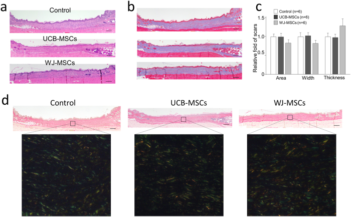

 Control group,
  UCB-MSC group,
  WJ-MSC group. *p < 0.05 vs. Control group. †p < 0.10 vs. Control group.
References
-
- Martin P. Wound healing–aiming for perfect skin regeneration. Science (New York, N.Y.) 276, 75–81 (1997). - PubMed
Publication types
MeSH terms
Substances
LinkOut - more resources
Full Text Sources
Other Literature Sources
Miscellaneous
