. 2016 Mar 11;291(11):5500-5511.
doi: 10.1074/jbc.M115.691162. Epub 2016 Jan 4.

Novel Interaction Mechanism of a Domain Antibody-based Inhibitor of Human Vascular Endothelial Growth Factor with Greater Potency than Ranibizumab and Bevacizumab and Improved Capacity over Aflibercept

Affiliations

Novel Interaction Mechanism of a Domain Antibody-based Inhibitor of Human Vascular Endothelial Growth Factor with Greater Potency than Ranibizumab and Bevacizumab and Improved Capacity over Aflibercept

Adam Walker et al. J Biol Chem. .

Abstract

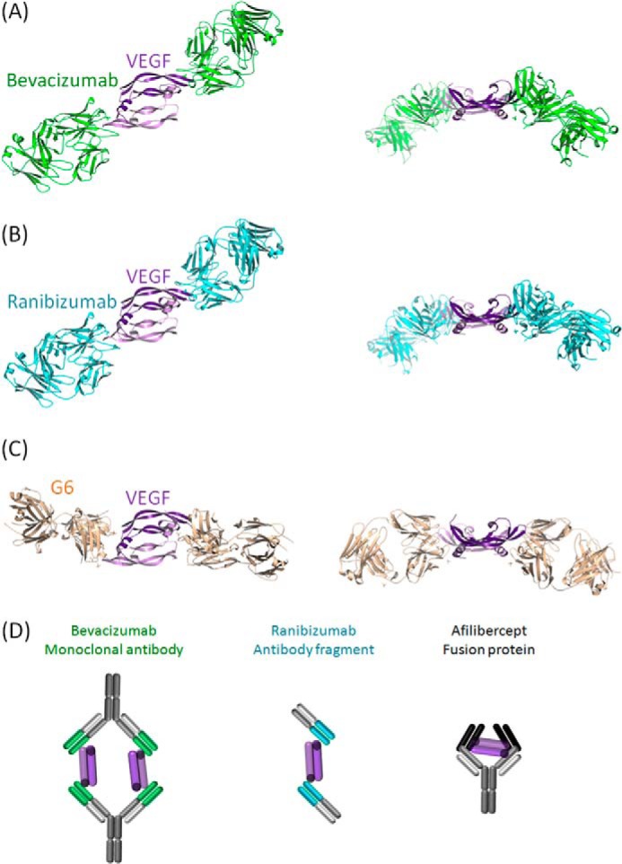
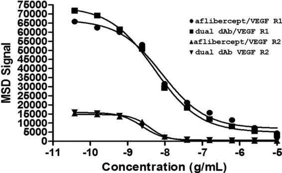
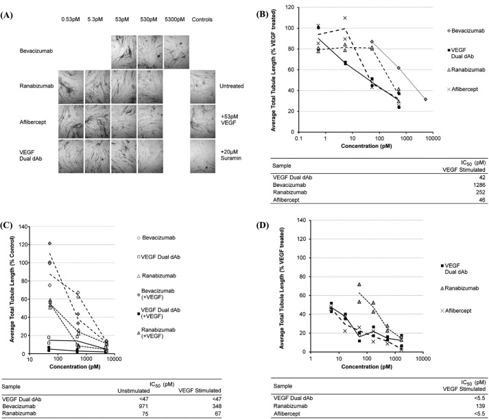
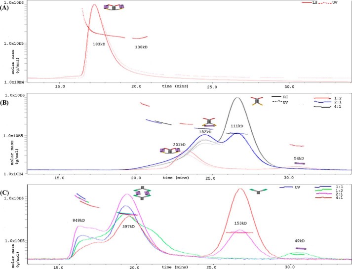
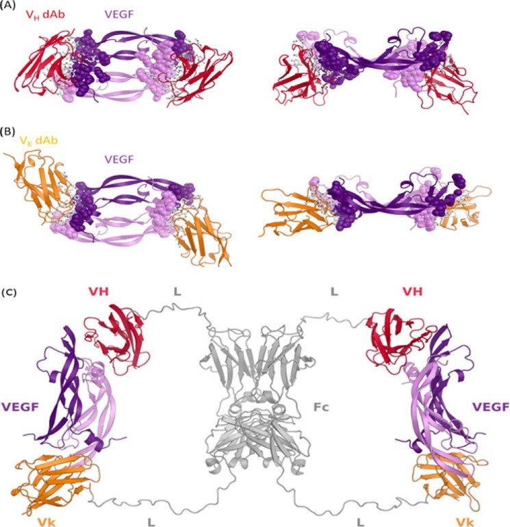
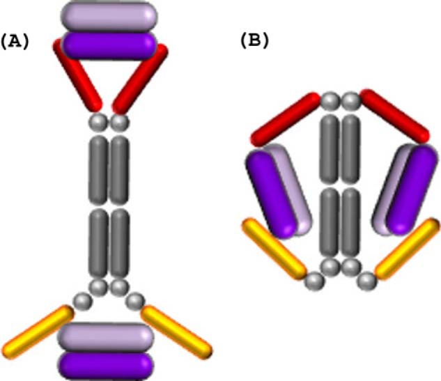
A potent VEGF inhibitor with novel antibody architecture and antigen binding mode has been developed. The molecule, hereafter referred to as VEGF dual dAb (domain antibody), was evaluated in vitro for binding to VEGF and for potency in VEGF-driven models and compared with other anti-VEGF biologics that have been used in ocular anti-angiogenic therapeutic regimes. VEGF dual dAb is more potent than bevacizumab and ranibizumab for VEGF binding, inhibition of VEGF receptor binding assays (RBAs), and VEGF-driven in vitro models of angiogenesis and displays comparable inhibition to aflibercept (Eylea). VEGF dual dAb is dimeric, and each monomer contains two distinct anti-VEGF domain antibodies attached via linkers to a human IgG1 Fc domain. Mechanistically, the enhanced in vitro potency of VEGF dual dAb, in comparison to other anti-VEGF biologics, can be explained by increased binding stoichiometry. A consistent model of the target engagement has been built based on the x-ray complexes of each of the two isolated domain antibodies with the VEGF antigen.

Keywords: angiogenesis; antibody engineering; drug action; protein-protein interaction; retinal degeneration; single-domain antibody (sdAb, nanobody); vascular endothelial growth factor (VEGF).


Figures

FIGURE 1.
FIGURE 1.
Experimental x-ray coordinates from the PDB for VEGF engagement of bevacizumab (Avastin, PDB code 1BJ1) (A), ranibizumab (Lucentis, PDB code 1CZ8) (B), and G6 antibody (PDB code 2FJG) (C) and schematic models of VEGF engagement for bevacizumab, ranibizumab, and aflibercept (Eylea/VEGF trap) (D) (34, 37, 38).
FIGURE 2.
FIGURE 2.
Activity of VEGF dual dAb and aflibercept in receptor binding assays. Inhibition of VEGF binding to VEGFR1 by aflibercept (●) or VEGF dual dAb (■) and VEGF binding to VEGFR2 by aflibercept (▴) or VEGF dual dAb (▾) was measured by MSD. Representative data are from one experiment (n = 3 similar experiments performed; see Table 4).
FIGURE 3.
FIGURE 3.
Inhibition of VEGF-driven tubule-formation in vitro by VEGF dual dAb, aflibercept, ranibizumab, and bevacizumab. A, representative tubule images from control and anti-VEGF-treated wells. Data were from one of three experiments; a single representative image of the three captured from each culture well using a 4× objective lens is shown. B, impact of anti-VEGF treatment on the total tubule length. Data were normalized to the 53 pm VEGF-treated positive control wells, and individual data points are plotted along with a line through the average value for each condition. Approximate IC50 values were determined from the inhibition curves. C, comparison of inhibitory activity of VEGF dual dAb, bevacizumab, and ranibizumab in untreated or VEGF-165 stimulated cultures, bevacizumab (♢) + VEGF (♦), VEGF dual dAb (□) + VEGF (■), and ranibizumab (▵) + VEGF (▴). D, comparison of inhibitory activity of VEGF dual dAb, aflibercept, and ranibizumab in VEGF-165-stimulated cultures, VEGF dual dAb + VEGF (■), ranibizumab + VEGF (▴), and aflibercept + VEGF (X). All conditions were tested in duplicate sample wells. Individual data points are plotted along with a line through the average value for each condition. Approximate IC50 values were determined from the inhibition curves.
FIGURE 4.
FIGURE 4.
SEC-MALLS analysis of anti-VEGF molecules with VEGF. Retention time (min) is shown on the x axis, and molecular mass (g/mol) is shown on the y axis. The line across the peak is the molecular weight distribution assigned by MALLS across the peak resolved by SEC. The solid, dash, and dotted lines are signals from UV, refractive index, and light scattering, (molar mass calculated from concentration and light scattering), respectively. A, dual dAb in a 1:1 molar ratio complex with human VEGF1–165. Analysis is on the TSK3000. B, molar ratios of VEGF dual dAb-VEGF complex at 1:2, 2:1, and 4:1 were analyzed on the Superdex TM200 SWXL. C, shows molar ratios of bevacizumab-VEGF complex at 1:2, 1:1, 2:1, and 4:1 which were also analyzed on the TM200 SWXL and the SEC-MALLS profile compared with that of the VEGF dual dAb. Schematic models of the molecular interactions assigned to the major peaks are also shown based on Figs. 1A, 5C, and Fig. 6.
FIGURE 5.
FIGURE 5.
X-ray crystal structure of the VEGF·dAb complexes and proposed structure of VEGF/VEGF dual dAb complex. VEGF/Vκ·dAb complex (A) and VEGF/VH·dAb complex (B) structures are depicted as secondary structure schematics with Vκ·dAbs colored in gold, VH·dAbs colored in red, and the VEGF homodimer colored in purple. In A and B, side chains of VEGF epitope and dAb paratope residues are depicted as spheres and sticks, respectively. C, in silico built model showing how a dual-dAb molecule could engage VEGF. Modeling was based on the crystal structures of the VEGF-dAb complexes. Fc homodimer and linker regions are colored gray. Fc-fused VH and Vκ·dAb regions are in red and gold, respectively (VH = VH·dAb; Vk = Vκ·dAb; Fc = hIgG Fc domain; L = peptide linker).
FIGURE 6.
FIGURE 6.
Schematic of potential engagement mechanisms of the VEGF dual dAb molecule with two VEGF dimers. A, end-on (where the two VH·dAbs fail to fully entrap the VEGF dimer). B, side-on; the VEGF dimer is displayed in purple, the VH·dAbs in red, the Vκ·dAbs in gold, and the IgG Fc backbone and linkers in gray.

References

    1. Freund K. B., Mrejen S., and Gallego-Pinazo R. (2013) An update on the pharmacotherapy of neovascular age-related macular degeneration. Expert Opin. Pharmacother. 14, 1017–1028 - PubMed
    1. Cheung N., Lam D. S., and Wong T. Y. (2012) Anti-vascular endothelial growth factor treatment for eye diseases. Brit Med. J. 344, e2970. - PubMed
    1. Kim Y. C., Chiang B., Wu X., and Prausnitz M. R. (2014) Ocular delivery of macromolecules. J. Control. Release 190, 172–181 - PMC - PubMed
    1. Yang J., Wang Q., Qiao C., Lin Z., Li X., Huang Y., Zhou T., Li Y., Shen B., Lv M., Feng J. (2014) Potent anti-angiogenesis and anti-tumor activity of a novel human anti-VEGF antibody, MIL60. Cell Mol. Immunol. 11, 285–293 - PMC - PubMed
    1. Tugues S., Koch S., Gualandi L., Li X., and Claesson-Welsh L. (2011) Vascular endothelial growth factors and receptors: anti-angiogenic therapy in the treatment of cancer. Mol. Aspects Med. 32, 88–111 - PubMed

MeSH terms