. 2016 Oct;21(10):1449-59.
doi: 10.1038/mp.2015.203. Epub 2016 Jan 5.

Early postnatal GABAA receptor modulation reverses deficits in neuronal maturation in a conditional neurodevelopmental mouse model of DISC1

Affiliations

Early postnatal GABAA receptor modulation reverses deficits in neuronal maturation in a conditional neurodevelopmental mouse model of DISC1

A Saito et al. Mol Psychiatry. 2016 Oct.

Abstract

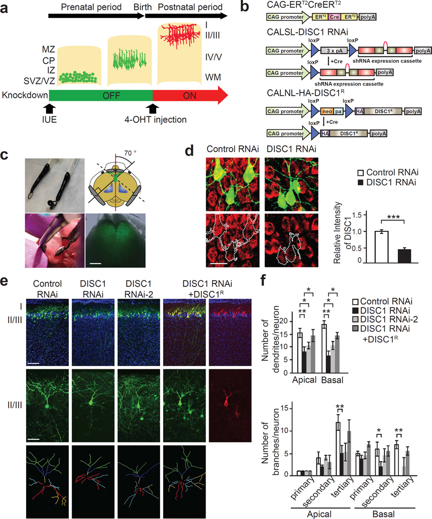
Exploring drug targets based on disease-associated molecular mechanisms during development is crucial for the generation of novel prevention and treatment strategies for neurodevelopmental psychiatric conditions. We report that prefrontal cortex (PFC)-specific postnatal knockdown of DISC1 via in utero electroporation combined with an inducible knockdown expression system drives deficits in synaptic GABAA function and dendritic development in pyramidal neurons, as well as abnormalities in sensorimotor gating, albeit without profound memory deficits. We show for the first time that DISC1 is specifically involved in regulating cell surface expression of α2 subunit-containing GABAA receptors in immature developing neurons, but not after full maturation. Notably, pharmacological intervention with α2/3 subtype-selective GABAA receptor positive allosteric modulators during the early postnatal period ameliorates dendritic deficits and behavioral abnormalities induced by knockdown of DISC1. These findings highlight a critical role of DISC1-mediated disruption of postnatal GABA signaling in aberrant PFC maturation and function.


Conflict of interest statement

A.C. and N.J.B. are full-time employees and shareholders in AstraZeneca. S.J.M serves as a consultant for SAGE therapeutics and AstraZeneca, relationships that are regulated by Tufts University.

Figures

Figure 1
Figure 1
Suppression of DISC1 expression impairs cell surface expression of GABAA receptor in developing neurons, but not in mature neurons. (a) Reduction of cell surface expression of GABAA receptor by knockdown of DISC1 (DISC1 RNAi) in developing cortical neurons at 7 days in vitro (7 DIV). Cell surface proteins were labeled by biotinylation, precipitated by avidin-conjugated beads, and immunoblotted for GABAA receptor α2, β2/3, and γ2 subunits. Cell surface expression of GABAA receptor α2, β2/3, and γ2 subunits (red arrowheads) was reduced by knockdown of DISC1 (left panels), but there was no change in their total expression (right panels). Reduction of their cell surface expression was reversed by co-expression of RNAi-resistant DISC1 (DISC1R). (b) Reduction of cell surface expression of GABAA receptors by immunocytochemistry with antibodies against α2, β3, and γ2 subunits (red traced by dotted white lines) under nonpermeant conditions in developing cortical neurons with knockdown of DISC1 (green) at 7 DIV. Blue, nucleus. Scale bar, 20 µm. (c) Knockdown of DISC1 has no effect on total or cell surface expression of GABAA receptors in mature cortical neurons at 30 DIV. TfR, transferrin receptor. (d) Impairment of GABAA receptor-dependent increase in [Ca2+] by knockdown of DISC1 in developing cortical neurons. Calcium imaging was assessed using the developing primary cortical neurons at 7 DIV transfected with GCaMP3 (green) as well as shRNAs to DISC1 (DISC1 RNAi) or scrambled controls (Control RNAi), which carry scarlet protein (red). Scale bar, 20 µm. (e) Bath application of GABA (50 µM) elicited increase in intracellular Ca2+ concentration in neurons with control RNAi, whereas knockdown of DISC1 impaired Ca2+ influx. Increase of calcium influx induced by GABA was prevented by treatment with bicuculline (10 µM). n = 10 neurons per condition. Bars represent averages of each group in three independent experiments (a, c). *P < 0.05, **P < 0.01, ***P < 0.001 determined by one-way ANOVA with posthoc Bonferroni test (a, e) and Student’s t test (c)
Figure 2
Figure 2
Postnatal suppression of DISC1 expression by selective targeting of pyramidal neurons in the prefrontal cortex (PFC) impairs dendritic development. (a) Schematic representation showing gene targeting by using the Cre/loxP system via in utero electroporation. Cells expressing inducible plasmids (green) that have migrated to the proper cortical layer; red cells represent post-migratory neurons, in which inducible plasmids were activated by intraperitoneal injection of 4-hydroxytamoxifen (4-OHT) during the postnatal period. MZ, marginal zone; CP, cortical plate; IZ, intermediate zone; SVZ/VZ, subventricular zone/ventricular zone. IUE, in utero electroporation. (b) 4-OHT activates Cre recombinase (Cre) comprising two mutated estrogen receptor (ER) binding regions of CAG-ERT2CreERT2 vector. Mir30-based shRNAs to DISC1 and HA-tagged RNAi-resistant DISC1 are expressed under control of the CAG promoter in the presence of 4-OHT-activated Cre (CALSL-DISC1 RNAi and CALNL-HA-DISC1R). (c) Three-electrode configuration for in utero electroporation to target the bilateral PFC. By using the custom-made electrode (upper panels), CALNL-GFP was simultaneously electroporated into the right and left side of PFC at embryonic day 14.5 (E14.5) and GFP expression was confirmed in the bilateral PFC at postnatal day 7 (P7) (lower panels). Scale bar, 1 mm. (d) Inducible DISC1 RNAi or Control RNAi were co-introduced with CAG-ERT2CreERT2 and CALNL-GFP in the PFC at E14.5 followed by intraperitoneal injection of 4-OHT at P2. Efficient suppression of DISC1 immunoreactivity (red traced by dotted white lines) in GFP-positive pyramidal neurons (green) is shown in the medial PFC at P7. n = 30 neurons from three mice per condition. Scale bar, 10 µm. (e, f) Mice with postnatal knockdown of DISC1 in pyramidal neurons of the medial PFC show impaired dendritic development, but not radial neuronal migration. Total number of dendrites and dendrite branches in each category (primary, secondary, and tertiary of apical and basal dendrites) was analyzed at P7. DISC1 RNAi has a stronger effect than RNAi-2. Inducible co-expression with RNAi-resistant DISC1 (red) normalized dendritic abnormalities induced by knockdown of DISC1. In each condition, 10 neurons from independent brain slices were analyzed in a blinded manner. Traced dendrites were categorized as primary apical (yellow) and basal (red), secondary apical (blue) and basal (cyan) dendrites, as well as tertiary apical (green) and basal (yellow) dendrites, respectively. Only neurons with typical morphological characteristics of pyramidal neurons (single thick axon arising from the basal side of the soma with multiple basal dendrites and apical dendrites extending vertically to layer I) were analyzed. Green, GFP; blue, nucleus. Scale bar, 100 µm (upper panels), 20 µm (middle panels). *P < 0.05, **P < 0.01, and ***P < 0.001 determined by Student’s t test (d) and one-way ANOVA with posthoc Bonferroni test (f).
Figure 3
Figure 3
Postnatal suppression of DISC1 expression in the prefrontal cortex (PFC) impairs developmental synaptic GABA function. (a) Immunohistochemical analysis with antibodies against GABAA receptor α2 subunits (red) and gephyrin, a GABA synaptic scaffold protein (green), in developing pyramidal neurons (blue) of the medial PFC in mice with postnatal knockdown of DISC1 at P14. Reduction of colocalization of GABAA receptor α2 subunits and gephyrin are observed in postnatal DISC1 knockdown mice. Scale bar, 10 µm. n = 5 neurons per condition. (b) Immunohistochemical analysis with antibodies against vesicular GABA transporter (vGAT, a presynaptic marker, indicated in red) and gephyrin (a GABA synaptic scaffold protein, indicated in green), in the developing pyramidal neurons (blue) of the medial PFC in mice with postnatal knockdown of DISC1 and control mice at P14. No changes in the number of gephyrin puncta and colocalization of vGAT and gephyrin are observed in postnatal DISC1 knockdown mice compared to control mice. Scale bar, 10 µm. n=5 neurons per condition. (c, d) Acute brain slice electrophysiology recordings from P14 mice show that postnatal suppression of DISC1 depresses GABAergic transmission. Representative traces depicting GABA-mediated miniature postsynaptic currents (GABA-mediated mPSCs) from each condition are shown. Summary graphs showing the amplitude of GABA-mediated mPSCs are decreased by postnatal DISC1 suppression, while DISC1 suppression does not affect on mPSCs frequency. All recordings were performed in the presence of D,L AP5 (100 µM), NBQX (10 µM), and TTX (1 µM). n = 10–15 neurons per condition. *P < 0.05 determined by Student’s t test (a, d).
Figure 4
Figure 4
Postnatal suppression of DISC1 expression in the prefrontal cortex (PFC) causes deficits in sensorimotor gating in young adulthood. (a) Postnatal knockdown of DISC1 in the PFC has no effect on locomotion as indicated by the open field test. (b) Postnatal suppression of DISC1 expression in the PFC has no effect on anxiety as indicated by the elevated plus maze test. (c) In the novel object recognition test, there were no differences in exploratory preference during training and retention sessions (left graph), and no abnormality in spatial recognition memory in postnatal DISC1 knockdown mice (right graph). (d) Excess acoustic startle response was observed in inducible DISC1 knockdown mice. n = 12 per condition. (e) Impairment of prepulse inhibition in the postnatal DISC1 suppression mice, when the 120 dB pulse was preceded by prepulses of 74, 78, and 82 dB. There were significant effects of genotype (F(1,110) = 53.92, P < 0.0001), prepulse intensity (F(4,110) = 29.93, P < 0.0001), and genotype-prepulse interaction (F(4,110) = 3.15, P = 0.0172). n = 12 per condition. (f) Projections from the PFC to nucleus accumbens core (NAc core), nucleus accumbens shell (NAc shell), and basolateral amygdala (BLA) were visualized by immunohistochemistry with antibodies against GFP (green). Postnatal DISC1 knockdown induced reductions in elevation of c-Fos expression (red) in the BLA, but not in the NAc core and NAc shell, following PPI. Blue, nucleus. Scale bar, 100 µm. n = 10 per condition. *P < 0.05, **P < 0.01, and ***P < 0.001 determined by Student’s t test (d, f) and two-way repeated measures ANOVA followed by the Bonferroni test (e).
Figure 5
Figure 5
Subtype-selective positive allosteric modulators of α2/3-containing GABAA receptors reverse dendritic abnormalities and altered behaviors induced by knockdown of DISC1. (a) Representative images showing effect of co-expressing RNAi-resistant DISC1 (DISC1R), and 7-day treatment with positive allosteric modulators of α2/3-containing GABAA receptors, AZD7325 (0.1 µM) and AZD6280 (0.1 µM), in primary cortical neuron expressing DISC1 RNAi carrying GFP protein (green). Traced dendrites were depicted as white lines (lower panels). Blue, nucleus. Scale bar, 100 µm. (b) Quantification of dendritic morphometry demonstrates that reduction of dendrite growth and branches as well as altered dendrite complexity induced by knockdown of DISC1 was ameliorated by co-expression with RNAi-resistant DISC1, in which the effect is comparable to treatment with AZD7325 and AZD6280. We analyzed only neurons with typical morphological characteristics of pyramidal neurons (single axon and multiple dendrites). n = 7 per condition. (c) Representative image showing dendritic structure of GFP-labeled pyramidal neurons in medial PFC from postnatal DISC1 suppression mice after intraperitoneal injection of AZD7325 (10 µmol/kg body weight) or vehicle for 1 week in the early postnatal period. Ratio of GFP fluorescence intensity in dendrites to total GFP fluorescence intensity was increased in AZD7325-treated inducible DISC1 knockdown mice, compared to those of vehicle-treated ones. Blue, nucleus. Scale bar, 100 µm. Ten brain slice images per condition were analyzed in a blinded manner. (d) Startle response to 120dB acoustic pulse was increased in the postnatal DISC1 knockdown mice compared to the control mice, and this excessive response was normalized by early postnatal treatment with AZD7325. (e) Impairment of prepulse inhibition in the postnatal DISC1 knockdown mice was rescued by AZD7325 treatment, significantly ameliorated at 74dB prepulse, and partially ameliorated at 78dB. *P < 0.05, **P < 0.01, and ***P < 0.001 determined by one-way ANOVA with posthoc Bonferroni test (b, c, d, e).

References

    1. Fritschy JM, Panzanelli P. GABAA receptors and plasticity of inhibitory neurotransmission in the central nervous system. Eur J Neurosci. 2014;39:1845–1865. - PubMed
    1. Ben-Ari Y. Excitatory actions of gaba during development: the nature of the nurture. Nat Rev Neurosci. 2002;3:728–739. - PubMed
    1. Cancedda L, Fiumelli H, Chen K, Poo MM. Excitatory GABA action is essential for morphological maturation of cortical neurons in vivo. J Neurosci. 2007;27:5224–5235. - PMC - PubMed
    1. Le Magueresse C, Monyer H. GABAergic interneurons shape the functional maturation of the cortex. Neuron. 2013;77:388–405. - PubMed
    1. Charych EI, Liu F, Moss SJ, Brandon NJ. GABA(A) receptors and their associated proteins: implications in the etiology and treatment of schizophrenia and related disorders. Neuropharmacology. 2009;57:481–495. - PMC - PubMed

Publication types

MeSH terms