. 2016 Jul;21(7):925-35.
doi: 10.1038/mp.2015.182. Epub 2016 Jan 5.

The autism-associated MET receptor tyrosine kinase engages early neuronal growth mechanism and controls glutamatergic circuits development in the forebrain

Affiliations

The autism-associated MET receptor tyrosine kinase engages early neuronal growth mechanism and controls glutamatergic circuits development in the forebrain

Y Peng et al. Mol Psychiatry. 2016 Jul.

Abstract

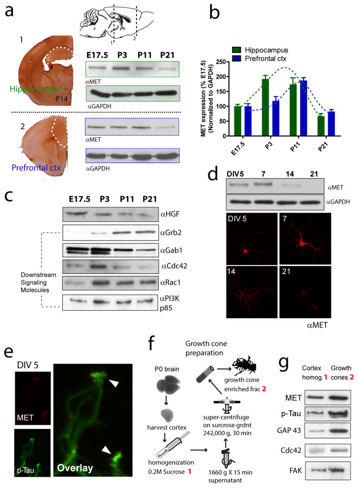
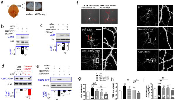
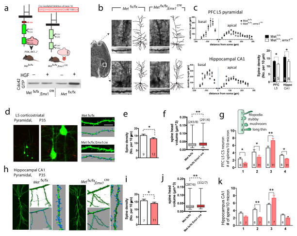
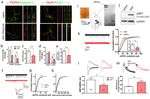
The human MET gene imparts a replicated risk for autism spectrum disorder (ASD), and is implicated in the structural and functional integrity of brain. MET encodes a receptor tyrosine kinase, MET, which has a pleiotropic role in embryogenesis and modifies a large number of neurodevelopmental events. Very little is known, however, on how MET signaling engages distinct cellular events to collectively affect brain development in ASD-relevant disease domains. Here, we show that MET protein expression is dynamically regulated and compartmentalized in developing neurons. MET is heavily expressed in neuronal growth cones at early developmental stages and its activation engages small GTPase Cdc42 to promote neuronal growth, dendritic arborization and spine formation. Genetic ablation of MET signaling in mouse dorsal pallium leads to altered neuronal morphology indicative of early functional maturation. In contrast, prolonged activation of MET represses the formation and functional maturation of glutamatergic synapses. Moreover, manipulating MET signaling levels in vivo in the developing prefrontal projection neurons disrupts the local circuit connectivity made onto these neurons. Therefore, normal time-delimited MET signaling is critical in regulating the timing of neuronal growth, glutamatergic synapse maturation and cortical circuit function. Dysregulated MET signaling may lead to pathological changes in forebrain maturation and connectivity, and thus contribute to the emergence of neurological symptoms associated with ASD.


Conflict of interest statement

CONFLICT OF INTEREST

The authors declare no conflict of interest.

Figures

Figure 1
Figure 1. Temporal profile of MET expression and growth cone enrichment in the developing forebrain
(a) IHC staining in P14 coronal slices reveals strong immunoreactivity in the hippocampus and PFC (delineated by white dotted lines). MET protein expression in the hippocampus and PFC was probed by Western blot analysis at E17.5, P3, P11 and P21. (b) Quantification of a. Peak expression of MET protein (normalized to GAPDH levels) occurs around P3-11, and declines precipitously thereafter. (c) Expression levels of the MET receptor ligand, HGF, and molecules potentially mediating downstream signaling events (Gab1, Grb2, Cdc42, Rac1 and PI3K/P85) (n = 2). (d) Western blot analysis (n = 3) and immunocytochemistry staining of MET protein in cultured low-density hippocampal neurons during in vitro development. (e) High resolution confocal imaging reveals strong MET immunoreactivity localized to axon growth cone tip (n = 4 experiments). (f) Diagram depicting isolation of growth cone fractions from P0 mouse brain. (g) Western blot analysis of MET reveals enriched protein levels at the growth cone, similar to other proteins known to be concentrated in growth cones (p-Tau, GAP43, Cdc42 and FAK) during early neuronal development (n = 3, quantification not shown).
Figure 2
Figure 2. MET signaling activates the small GTPase Cdc42, which mediates its effects on dendritic and spine morphogenesis
(a) Schematic illustration of an ex vivo prefrontal brain slice preparation (coronal sections from P9 mice) used for HGF treatment to initiate MET signaling. (b) MET activation, using p-MET (Y1234/1235) level as a surrogate, was induced by HGF, and blocked by MET kinase activity inhibitor PHA665752 (200 nM) (**p < 0.01. ND, not detected). (c) The PI3K inhibitor, Wortmanin (100 nM), did not affect p-MET levels, indicating PI3K activity was not required MET activation. (d) In both P9-10 ex vivo slices and DIV11 cortical neuron culture preparations, HGF (50 ng/ml) induces Cdc42 activation, measured by PAK P21 domain pull-down of the GTP-bound Cdc42. In contrast, no change of overall Cdc42 level was observed (*p < 0.05, **p < 0.01). (e) Cdc42 activation was blocked by either PHA665752 or Wortmanin, indicating it is dependent on MET kinase activity and downstream to PI3K activation (**p < 0.01). (f) Neuronal morphometric measurement using TDBTN and TDBL, and representative images of dendritic and spine morphology from cultured neurons transfected with Met cDNA, or in combination with Cdc42 loss-of -function plasmids (DN-Cdc42 or Cdc42 RNAi). (g) TDBTN quantification (normalized to GFP control neurons) reveals MET OE significantly increased TDBTN, which is significantly blocked by either DN-Cdc42 or Cdc42 RNAi co-transfection. Note Cdc42 RNAi alone significantly reduced TDBTN (*p < 0.05, **p < 0.01 compared with GFP, ns, not significant. ##p < 0.01. Number of neurons quantified listed on bar). (h) MET OE increases TDBL, an effect that is also blocked by co-transfection with DN-Cdc42 or Cdc42 RNAi (*p < 0.05, **p < 0.01, #p < 0.05, ##p < 0.01). (i) Enhanced MET signaling increases dendritic spine density, which is blocked by Cdc42 antagonism (**p < 0.01, ##p < 0.01, ns, not significant).
Figure 3
Figure 3. Conditional Met knockout leads to morphological alterations in dendritic and spine structures in forebrain neurons
(a) Schematic representation of dorsal pallium specific Met cKO (Met fx/fx; emx1cre) mice. Cre-mediated excision of Exon 16, which encodes a critical ATP-binding site (Lys1108), inactivates the intracellular tyrosine kinase activity of MET and abrogates adaptor protein recruitment and downstream signaling. Consistently, HGF treatment in cKO PFC slices failed to induce an increase in pY1234/1235 (n = 2, quantification not shown). (b) Photomicrograph of Golgi-stained prefrontal (infra-limbic) L5 pyramidal neuron, and hippocampus CA1 neuron in Metfx/fx and cKO brains. A representative dendritic arbor reconstructed in 3D using Neurolucida for each neuron was shown on the right, and dendritic segments from which dendritic spine density was calculated were shown below the image. (c) Sholl analysis of PFC L5 and hippocampal CA1 neuron dendritic complexity. A significant reduction of dendritic length was found in PFC L5 (two-way ANOVA with Bonferroni post hoc test, F(1, 288) = 19.06. n = 7 neurons/3 mice for Metfx/fx and n = 8/4 for cKO. *p < 0.05). No significant overall difference was found for CA1 neurons (F(1, 315) = 0.003. n = 8 neurons/3 mice for Metfx/fx and n = 9/3 for cKO), while dendritic length was distributed differently (*p < 0.05). Dendritic spine density showed a significant reduction in both prefrontal L5 pyramidal neuron and CA1 pyramidal neuron in cKO brains (n = 10 neurons/3 mice, *p < 0.05, t test). (d) Dendritic spine morphology in P35 PFC L5 corticostriatal neurons, labeled by red retrograde ‘beads’, was reconstructed using Imaris and high-resolution confocal Z stack images. (e) Met cKO PFC L5 neurons showed a significant reduction in spine density (t19 = 3.67, * p < 0.05). (f) Dendritic spine head volume in cKO PFC L5 neurons is significantly larger (**p < 0.01). (g) Dendritic spines from cKO PFC L5 neurons show significantly increased number in ‘mushroom’ type (**p < 0.01), while significant decreased numbers in all other categories (* p < 0.05). Numbers labeled on bar denotes number of neurons, based on which > 500 spines were analyzed for each group. (h) Morphological reconstruction of CA1 neuron spines. (i–j) CA1 neurons from cKO mice show a significant reduction in spine density (i, t16 = 3.42, *p < 0.05), and a significant increase in dendritic spine head volume (j, **p < 0.01). (k) CA1 neuron dendritic spines from cKO mice show significant increased number in ‘mushroom’ type (**p < 0.01), and significant decreased numbers in the ‘stubby’ category (**p < 0.01).
Figure 4
Figure 4. Prolonged HGF/MET signaling reduces functional glutamatergic synapse formation and represses its maturation
(a) Representative images of immunocytochemistry staining of DIV25 cultured cortical neurons with or without HGF treatment. Two pairs of synapse markers, the post-synaptic protein PSD95 / presynaptic protein synapsin I; and the NMDA receptor subunit GluN1 / AMPA receptor subunit GluA1 were used to estimate functional synapse (colocalized puncta in yellow). (b) Quantification of staining in a. Neurons from HGF treated groups show significantly reduced puncta density in both PSD95 and synapsin I staining (**p < 0.01). HGF also significantly reduced the proportion of putative functional synapse number, defined by PSD95+/Synapsin I+ puncta (c. t25 = 7.73, ***p < 0.001). (d) HGF-treated neurons display reduced GluA1+ puncta density (t17 = 4.33, **p < 0.01). (e) HGF also significantly reduced the proportion of putative functional synapse number, defined by GluN1+/GluA1+ puncta (t19 = 7.84, ***p < 0.001). (f) Representative mEPSC recording from a control- (saline) and HGF-treated neuron. Below, averaged mEPSC from a 1-min epoch was superimposed for comparison. (g) Compared with saline-treated neurons, HGF-treated neurons displayed significantly reduced mEPSC amplitude distribution (n > 2500 events/6 neurons for both groups, **p < 0.01, K-S test) and increased inter-event intervals (h, n > 2000 events/8 neurons, *p < 0.05, K-S test). (i) Illustration of cultured brain slices containing the PFC region, with a L5 neuron targeted for whole cell patch clamp recording, and L2/3 was electrically stimulated to obtain monosynaptic responses. (j) HGF treatment for in cultured slices leads to prolonged MET activation, measured by increased pY1234/1235 (n = 2, quantification not shown). (k) Representative mEPSC recording from a L5 PFC neuron in control (saline) and HGF-treated cultured slices. Quantification reveals that HGF-treatment significantly reduced mEPSC amplitude (n > 3000 events/9 neurons for each group, ***p < 0.001, K-S test) and frequency (t19 = 5.21, **p < 0.01). (l) HGF-treated L5 neurons showed significantly reduced A:N current ratio (t20 = 3.89, *p < 0.05) compared with saline-treated control neurons. (m) HGF-treated L5 neurons showed significantly increased blockade of NMDAR current by ifenprodil (t13 = 4.21, **p < 0.01) compared with saline treated control neurons.
Figure 5
Figure 5. Single neuron alternation in MET signaling during in vivo development disrupts local cortical circuit connectivity
(a) Experimental paradigm. C57Bl6 embryos were electroporated with MET OE, RNAi plasmids in combination with GFP, or control GFP alone at E13.5. A DIC image of live coronal slice superimposed with the GFP channel was shown to denote PFC L5 neuron targeting by IUEP. (b) Illustration of a sagittal PFC slice prepared from IUEP mice, from which L5 PFC neurons were selected for LSPS mapping. A stimulation grid of 16*16 was overlaid to denote stimulation locations. Glutamate uncaging using UV laser pulses produces three types of responses, a direct response (red trace) when glutamate was uncaged at a location close to the soma or dendrites; a synaptic circuit response (green) when glutamate was uncaged and activated a synaptically connected presynaptic neuronal population; or an inhibitory response (voltage clamped at 0 mV) in the form of outward current from synaptically connected interneurons. (c) An example of a matrix of excitatory synaptic input responses to a L5 PFC neuron electroporated with GFP, in response to glutamate uncaging at locations indicated in b. Note direct responses were not plotted. The pseudo-color representation (i.e. ‘map’) of the location and strength of these responses was shown to the right. (d) An inhibitory response map was collected at 0 mV holding potential following the excitatory map. (e) Averaged excitatory (n = 12 neurons/4 mice) maps from L5 neurons with GFP electroporation. For each individual map, the 16 columns of responses were binned into L2/3, L5A, L5B and L6, and the averaged EPSC and IPSC was calculated for each column. Mean and standard errors of the pooled maps were plotted to the right of the average map. Averaged inhibitory inputs (n = 10/4) and their laminar distribution were shown in the panel below. (f) Averaged excitatory (top panel, n = 19/3) and inhibitory (n = 8/3) maps for L5 PFC neurons electroporated with Met cDNA. (g) Averaged excitatory (top panel, n = 13/4) and inhibitory (n = 9/4) maps for L5 PFC neurons electroporated with MET RNAi. (h) Pooled excitatory synaptic inputs to all groups of L5 neurons as a function of their laminar location. Two-way ANOVA revealed a significant treatment effect (F(2, 656) = 15.8), with significantly increased input strength in MET OE neurons at L5A and L5B (*p < 0.05, Bonferroni post hoc test). In contrast, RNAi neurons showed significantly reduced synaptic inputs from L2/3 (#p < 0.05, Bonferroni post hoc test). (i) Pooled inhibitory synaptic inputs to all groups of L5 neurons as a function of their laminar location. No significant difference was detected for the treatment effects on laminar distribution of inhibitory inputs (F(2, 384) = 0.09. p > 0.05).

References

    1. Park H, Poo MM. Neurotrophin regulation of neural circuit development and function. Nat Rev Neurosci. 2013;14(1):7–23. - PubMed
    1. Naldini L, Vigna E, Ferracini R, Longati P, Gandino L, Prat M, et al. The tyrosine kinase encoded by the MET proto-oncogene is activated by autophosphorylation. Mol Cell Biol. 1991;11(4):1793–1803. - PMC - PubMed
    1. Uehara Y, Minowa O, Mori C, Shiota K, Kuno J, Noda T, et al. Placental defect and embryonic lethality in mice lacking hepatocyte growth factor/scatter factor. Nature. 1995;373(6516):702–705. - PubMed
    1. Judson MC, Amaral DG, Levitt P. Conserved subcortical and divergent cortical expression of proteins encoded by orthologs of the autism risk gene MET. Cereb Cortex. 2011;21(7):1613–1626. - PMC - PubMed
    1. Judson MC, Bergman MY, Campbell DB, Eagleson KL, Levitt P. Dynamic gene and protein expression patterns of the autism-associated met receptor tyrosine kinase in the developing mouse forebrain. J Comp Neurol. 2009;513(5):511–531. - PMC - PubMed

Publication types

Substances