. 2016 Jan 4;212(1):29-38.
doi: 10.1083/jcb.201507122.

PML isoform II plays a critical role in nuclear lipid droplet formation

Affiliations

PML isoform II plays a critical role in nuclear lipid droplet formation

Yuki Ohsaki et al. J Cell Biol. .

Abstract

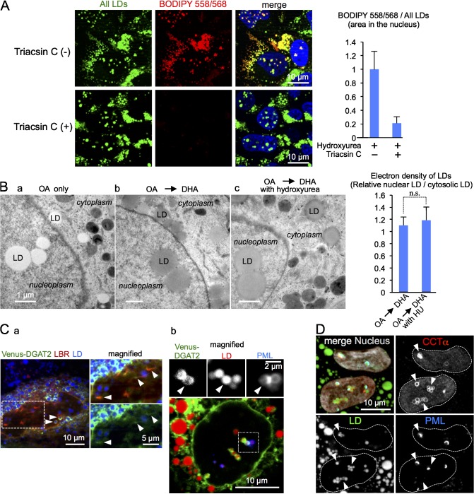
Lipid droplets (LDs) in the nucleus of hepatocyte-derived cell lines were found to be associated with premyelocytic leukemia (PML) nuclear bodies (NBs) and type I nucleoplasmic reticulum (NR) or the extension of the inner nuclear membrane. Knockdown of PML isoform II (PML-II) caused a significant decrease in both nuclear LDs and type I NR, whereas overexpression of PML-II increased both. Notably, these effects were evident only in limited types of cells, in which a moderate number of nuclear LDs exist intrinsically, and PML-II was targeted not only at PML NBs, but also at the nuclear envelope, excluding lamins and SUN proteins. Knockdown of SUN proteins induced a significant increase in the type I NR and nuclear LDs, but these effects were cancelled by simultaneous knockdown of PML-II. Nuclear LDs harbored diacylglycerol O-acyltransferase 2 and CTP:phosphocholine cytidylyltransferase α and incorporated newly synthesized lipid esters. These results corroborated that PML-II plays a critical role in generating nuclear LDs in specific cell types.


Figures

Figure 1.
Figure 1.
Nuclear LDs in Huh7. Cells used in this and subsequent figures were cultured with 0.4 mM OA for 12 h if not otherwise described. (A) Scheme of type I and type II NR. (B) Serial ultrathin sections (60 nm thick) of cells cultured with 0.2 mM DHA for 2 h to enhance the contrast of membranes. The LD indicated by the arrowhead was verified to be in the nucleoplasm. (C) Immunolabeling of perilipin2 (ADRP), perilipin3 (TIP47), and Rab18. Cytoplasmic LDs were labeled for all three, whereas nuclear LDs were labeled for perilipin3 and Rab18 (arrowheads), but not for perilipin2. Proteins (red), LDs (green), nucleus (blue). The bar graph shows the ratio of labeled LDs in the nucleus and the cytoplasm in one representative experiment out of three (mean ± SEM, n ≥ 99 cells/experiment). (D) Cytoplasmic LD counts were significantly lower in clones deleted with the perilipin2 gene than in the control, but nuclear LD counts were at similar levels. Two independent clones (1 and 2) were examined. The bar graph shows mean ± SD of pooled data from three independent experiments. n ≥ 100 cells/experiment. (E) Cells expressing HRP-KDEL were processed by DAB histochemistry to delineate the ER and the nuclear envelope. Nuclear LDs showed a close association with DAB precipitates, either in linear shapes (arrowheads) or in puncta (arrows), which were thought to represent the type I NR sectioned longitudinally or vertically, respectively. See Fig. S2 A for more examples. (F) Type I NR in association with nuclear LDs (arrowhead) was observed in cells without HRP-KDEL expression by conventional EM.
Figure 2.
Figure 2.
Nuclear LDs were associated with PML NBs in Huh7. (A) Colocalization of endogenous PML (red) and nuclear LDs (green; arrowheads). More than 70% of PML and >30% of nuclear LDs were associated with the other in cells cultured with OA for 12 h; the colocalization ratio per LDs increased when the number of nuclear LDs decreased in a lipoprotein-deficient medium. The bar graph shows mean ± SEM of one representative experiment out of three. n = 88 cells. (B) Pre-embedding immuno-EM. PML labeling was observed adjacent to nuclear LDs. (C) Correlative light and EM. Nuclear LDs that harbor EGFP-tagged PML-I showed a radiating bristle-like structure on the surface (arrowheads). (D) Distribution of PML isoforms (red). siRNA-resistant mCherry-PML isoform cDNAs were transfected to cells depleted of endogenous PML. Only PML-II showed prominent colocalization with nuclear LDs (green; arrowheads). The bar graphs (C–E) show mean ± SD of pooled data from three independent experiments. n ≥ 100 cells/experiment. (E) Overexpression of PML-II, but not of other isoforms, induced a significant increase in nuclear LDs compared with the empty vector control. (F) Knockdown of PML-II decreased nuclear LDs. Two siRNAs targeted at different sequences of PML-II (1 and 2) were used.
Figure 3.
Figure 3.
Correlation of the PML-II patch and nuclear LDs in Huh7. (A) Distribution of PML-II truncation mutants in cells depleted of endogenous PML-II. PML-II (FL, full length) and PML-II (1–716) showed colocalization with nuclear LDs (green; arrowheads) and linear distribution in the nuclear periphery (arrows), whereas PML-II (1–652) did not show either. (B) PML-II (FL) and PML-II (1–716), but not PML-II (1–652), induced a significant increase in nuclear LDs. The bar graph shows mean ± SD of pooled data from three independent experiments. n ≥ 100 cells/experiment. (C) The PML-II patch and nuclear LDs formed concomitantly in a cell type–specific manner. mCherry-PML-II was targeted at both LD-associated PML NBs (arrowheads) and the nuclear periphery (arrows) in Huh7 and U2OS, but mCherry-PML-II distributed only to PML NBs in Y1 and HeLa. (D) Pre-embedding immuno-EM of GFP–PML-II. The labeling was observed on the nucleoplasmic side of the INM. ONM, outer nuclear membrane. (E) LBR and SUN1 were excluded from the PML-II patch region of the nuclear envelope (arrowheads).
Figure 4.
Figure 4.
Correlation of PML-II, nuclear LDs, and intranuclear membranes. (A) Knockdown of SUN2 alone or both SUN1 and SUN2 in Huh7 caused increases in nuclear LDs (green) and intranuclear membranes labeled for LBR (red), but the increases were cancelled by additional knockdown of PML-II. In this experiment, OA was not added to the culture medium. The bar graph shows mean ± SD of pooled data from three independent experiments. n ≥ 100 cells/experiment. (B) Type I NR observed in Huh7 after SUN2 knockdown. See Fig. S3 C for more examples. (C) Overexpression of PML-II in Huh7 significantly increased intranuclear membranes labeled for LBR (arrowheads). The bar graph shows mean ± SD of pooled data from three independent experiments. n ≥ 100 cells/experiment. (D) Knockdown of REEP3/4 in Huh7 significantly increased nuclear LDs (green) and intranuclear membranes (red), but the increases were not observed when both REEP3/4 and PML-II were knocked down. Cells were cultured without OA. (E) Correlation between PML-II and nuclear LDs. In most cell types, LDs and membranes are removed from chromatin or chromosome during mitosis by the mechanism involving SUN and REEP proteins. In the limited types of cells forming the PML-II patch (e.g., Huh7), LDs and membranes are potently tethered to chromatin by PML-II, leading to their frequent presence in the interphase nucleus. As shown in Fig. 5, lipid esters are thought to be synthesized in these intranuclear LDs and membranes.
Figure 5.
Figure 5.
Nuclear LDs in Huh7 grow by incorporating newly synthesized lipid esters. (A) Fluorescence microscopic assay. Cells were incubated for 1 h with a fluorescence fatty acid analog BODIPY558/568-C12 (red) and chased for another hour, all in the presence of 1.5 mM hydroxyurea to inhibit mitosis. After fixation, LDs were labeled with BODIPY493/503 (green; top). BODIPY558/568-positive LDs did not appear when treated with 5 µM triacsin C (bottom), which confirmed that the analog was incorporated into LDs as lipid esters. The bar graph shows mean ± SD of pooled data from three independent experiments. n ≥ 100 cells/experiment. (B) EM assay. Cells were cultured with either 0.4 mM OA for 17 h (a) or 0.4 mM OA for 9 h and then with 0.2 mM DHA for 8 h (b and c), without (b) or with (c) the presence of 1.5 mM hydroxyurea. The electron density of nuclear LDs was increased by DHA to the same degree as that of cytoplasmic LDs, irrespective of the hydroxyurea treatment. The bar graph shows mean ± SD of pooled data from four independent experiments. n = 156 (cytoplasmic LD) and n = 216 (nuclear LD). (C) DGAT2 in the nucleus. Venus-DGAT2 (green) showed (a) colocalization with RFP-LBR (red) and (b) distribution around nuclear LDs (red; arrowheads). Some of those LDs were positively labeled for PML (blue), which confirmed their presence in the nucleoplasm. (D) CCTα in the nucleus. Endogenous CCTα (red) was labeled around nuclear LDs (green; arrowheads), some of which were also labeled positively for PML (blue). CCTα-EGFP showed a similar distribution (Fig. S3 D).

Comment in

  • Lipid droplets go nuclear.
    Farese RV Jr, Walther TC. Farese RV Jr, et al. J Cell Biol. 2016 Jan 4;212(1):7-8. doi: 10.1083/jcb.201512056. J Cell Biol. 2016. PMID: 26728852 Free PMC article.

References

    1. Bernardi R., and Pandolfi P.P.. 2007. Structure, dynamics and functions of promyelocytic leukaemia nuclear bodies. Nat. Rev. Mol. Cell Biol. 8:1006–1016. 10.1038/nrm2277 - DOI - PubMed
    1. Berscheminski J., Groitl P., Dobner T., Wimmer P., and Schreiner S.. 2013. The adenoviral oncogene E1A-13S interacts with a specific isoform of the tumor suppressor PML to enhance viral transcription. J. Virol. 87:965–977. 10.1128/JVI.02023-12 - DOI - PMC - PubMed
    1. Bickel P.E., Tansey J.T., and Welte M.A.. 2009. PAT proteins, an ancient family of lipid droplet proteins that regulate cellular lipid stores. Biochim. Biophys. Acta. 1791:419–440. 10.1016/j.bbalip.2009.04.002 - DOI - PMC - PubMed
    1. Brasaemle D.L., Barber T., Kimmel A.R., and Londos C.. 1997. Post-translational regulation of perilipin expression. Stabilization by stored intracellular neutral lipids. J. Biol. Chem. 272:9378–9387. 10.1074/jbc.272.14.9378 - DOI - PubMed
    1. Brown W.J., and Farquhar M.G.. 1989. Immunoperoxidase methods for the localization of antigens in cultured cells and tissue sections by electron microscopy. Methods Cell Biol. 31:553–569. 10.1016/S0091-679X(08)61626-X - DOI - PubMed

Publication types

Associated data