. 2016 Jan 4:74:1.26.1-1.26.27.
doi: 10.1002/0471142301.ns0126s74.

Anterograde or Retrograde Transsynaptic Circuit Tracing in Vertebrates with Vesicular Stomatitis Virus Vectors

Affiliations

Anterograde or Retrograde Transsynaptic Circuit Tracing in Vertebrates with Vesicular Stomatitis Virus Vectors

Kevin T Beier et al. Curr Protoc Neurosci. .

Abstract

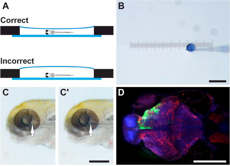
Viruses have been used as transsynaptic tracers, allowing one to map the inputs and outputs of neuronal populations, due to their ability to replicate in neurons and transmit in vivo only across synaptically connected cells. To date, their use has been largely restricted to mammals. In order to explore the use of such viruses in an expanded host range, we tested the transsynaptic tracing ability of recombinant vesicular stomatitis virus (rVSV) vectors in a variety of organisms. Successful infection and gene expression were achieved in a wide range of organisms, including vertebrate and invertebrate model organisms. Moreover, rVSV enabled transsynaptic tracing of neural circuitry in predictable directions dictated by the viral envelope glycoprotein (G), derived from either VSV or rabies virus (RABV). Anterograde and retrograde labeling, from initial infection and/or viral replication and transmission, was observed in Old and New World monkeys, seahorses, jellyfish, zebrafish, chickens, and mice. These vectors are widely applicable for gene delivery, afferent tract tracing, and/or directional connectivity mapping. Here, we detail the use of these vectors and provide protocols for propagating virus, changing the surface glycoprotein, and infecting multiple organisms using several injection strategies.

Keywords: VSV; axon tracing; gene delivery; neural circuitry; transsynaptic tracing.


Figures

Figure 1.26.1
Figure 1.26.1. Schematic overview of rVSV propagation procedures
Illustrations of (A) expansion of replication-competent rVSV stocks, (B) passage of replication-conditional vectors, and (C) generation of rVSVΔG(EnvA) with minimal contamination of the previous G protein.
Figure 1.26.2
Figure 1.26.2. Cytopathic effect (CPE) in 293T cells infected with replication-conditional and replication-competent rVSV stocks
Example images of (A–C) uninfected control cells, (D–F) rVSVΔG(VSV-G) expressing tdTomato, and fluorescence from (G–I) rVSV(VSV-G) expressing Venus, (J–L) rVSVΔG(RABV-G) expressing GFP, and (M–O) rVSV(RABV-G) expressing GFP infected 293T cells at 20 hr post infection (hpi). Morphological rounding of cells is observed in infected 293T cells (white arrows).
Figure 1.26.3
Figure 1.26.3. Serial dilution of rVSV for viral titration
Step 1: Pipet 2 μl of concentrated stock into the first well. Step 2: Pipet 2 μl of concentrated stock into tube containing 18 μl of DMEM (to make 1/10 dilution) and vortex to mix well. Step 3 and subsequent steps: To prepare serial 10-fold dilutions, pipet 2 μl of previous 20-μl dilution into the next 18-μl aliquot, and repeat until you have a dilution series at 10-fold increments (dilution range = 10−1 to 10−11). Step 4: Add 2 μl of an aliquot to a designated well, such that each well represents a different viral dilution. The number of infected cells per well can be counted at a later time point to determine viral titer.
Figure 1.26.4
Figure 1.26.4. Injection of rVSV into adult mice
After the mouse is anesthetized, place mouse in stereotaxic apparatus, (A) inserting the teeth into the bite bar, and securing the ear bars. (B) To ensure that the mouse is properly anesthetized, gently but firmly pinch the toes of the mouse. If it does not respond, proceed to the next steps. (C) Use a scalpel to make an incision along the top of the scalp. (D) Apply ophthalmic ointment to the eyes to prevent drying of the corneas. Move the injection pump to the desired coordinates, and drill a hole in the skull at the desired anterior/posterior and lateral/medial coordinates along the skull. After the hole has been drilled, (E) insert injection capillary through the hole, and stop at the proper dorsal/ventral coordinates. Inject the rVSV at a rate of 100 nl/min. After waiting at least 5 minutes, slowly withdraw the injection capillary. (F) When complete, remove the mouse from the stereotaxic apparatus, and suture the skin. Place the mouse on a heating pad to assist in recovery from anesthesia, providing analgesic administration as necessary. (G) An example brain, fixed 2 days post-injection, after a bilateral injection of rVSV(VSV-G) expressing YFP. (H) A coronal section of the brain shown in (G), showing infection of primary hippocampal neurons. (I) A parasagittal brain section indicating anterograde transsynaptic transmission of rVSV(VSV-G) after injection into the dorsal striatum. Shown is a section taken 3 days post-infection.
Figure 1.26.5
Figure 1.26.5. Injection of rVSV in chicken embryos
(A–C) To prevent lethality associated with injection at late stages, embryos and extraembryonic membranes must be lowered away from the shell at E2, prior to extensive vascularization. Use a syringe needle to make a pilot hole (A), prior to removal of 3–4 ml of albumin (B). Seal the hole by placing tape over the top surface of the egg (C). This will also prevent cracking of the egg during egg windowing 12 days later. (D–F) Injection of rVSV at E14. To window the egg, cut a circular opening through the tape and the top of the shell (D). Avoiding vasculature, make a small opening in the chorioallantoic membrane and grasp the beak of the embryo with forceps (E). Raise head and position the site to be injected against the overlaying membrane, away from major blood vessels. While maintaining position of the head, inject rVSV with a beveled tip Hamilton syringe into the desired location. Shown is an injection into the vitreous cavity of the right eye (F). (G–I) Anticipated results. Representative images of brightfield (G) and GFP expression (green) (G′) in a dissected retina, two days after rVSV(VSV-G) injection into the vitreous cavity of the eye. A representative section through the midbrain shows distinct GFP expression within the optic tectum (H–I). As expected, GFP-positive neurons are observed in the outer stratum griseum et fibrosum superficiale (SGFS) and the inner stratum griseum central (SGC) layers of the optic tectum (H–I), suggesting efficient rVSV transmission and labeling of visual circuits from the eye to the brain. Sections are counterstained with DAPI in H (blue).
Figure 1.26.6
Figure 1.26.6. Zebrafish injection setup
(A) Pneumatic pump, (B) stereomicroscope, (C) micromanipulator, and (D) forceps and pipette pump.
Figure 1.26.7
Figure 1.26.7. Zebrafish mounting and injection
(A) Zebrafish larvae are mounted in the center chamber of a glass-bottom dish (side view). Low-melting point agarose should cover the entire center chamber. However, an excessive amount of agarose will make needle positioning more difficult. (B) A droplet of virus mixed with Fast Green dye after a single pressure injection, with a diameter of ~100 μm. (C) For retinal injection, the glass pipette tip (arrow) is inserted into the vitreous cavity of the eye. After injection, the blue/green virus solution is visible in the retina and the eye becomes slightly swollen (C′). (D) Representative image of zebrafish injected with rVSV(VSV-G) expressing Venus, fixed 1 dpi. Shown here is a horizontal optical section, with the rostral direction facing left and caudal direction facing right. Injection of the left eye results in Venus labeling (green) in the contralateral (top) half of the brain. This sample is counterstained with anti-GABA (red) and anti-ERK (blue) antibodies. Scale bars = 200 μm.

References

    1. Baer A, Kehn-Hall K. Viral Concentration Determination Through Plaque Assays: Using Traditional and Novel Overlay Systems. Journal of Visualized Experiments. 2014;(93):1–10. doi: 10.3791/52065. - DOI - PMC - PubMed
    1. Beier KT, Borghuis BG, El-Danaf RN, Huberman AD, Demb JB, Cepko CL. Transsynaptic tracing with vesicular stomatitis virus reveals novel retinal circuitry. The Journal of Neuroscience. 2013;33(1):35–51. doi: 10.1523/JNEUROSCI.0245-12.2013. - DOI - PMC - PubMed
    1. Beier KT, Saunders AB, Oldenburg IA, Sabatini BL, Cepko CL. Vesicular stomatitis virus with the rabies virus glycoprotein directs retrograde transsynaptic transport among neurons in vivo. Frontiers in Neural Circuits. 2013;7:11. doi: 10.3389/fncir.2013.00011. - DOI - PMC - PubMed
    1. Beier KT, Saunders A, Oldenburg Ia, Miyamichi K, Akhtar N, Luo L, Cepko CL. Anterograde or retrograde transsynaptic labeling of CNS neurons with vesicular stomatitis virus vectors. PNAS. 2011;108(37):15414–15419. doi: 10.1073/pnas.1110854108. - DOI - PMC - PubMed
    1. Boussif O, Lezoualc’h F, Zanta Ma, Mergny MD, Scherman D, Demeneix B, Behr JP. A versatile vector for gene and oligonucleotide transfer into cells in culture and in vivo: polyethylenimine. PNAS. 1995;92(16):7297–7301. doi: 10.1073/pnas.92.16.7297. - DOI - PMC - PubMed

Publication types

Substances