Case Reports
. 2016 Jun;31(6):1200-6.
doi: 10.1002/jbmr.2778. Epub 2016 Feb 6.

A G-protein Subunit-α11 Loss-of-Function Mutation, Thr54Met, Causes Familial Hypocalciuric Hypercalcemia Type 2 (FHH2)

Affiliations
Case Reports

A G-protein Subunit-α11 Loss-of-Function Mutation, Thr54Met, Causes Familial Hypocalciuric Hypercalcemia Type 2 (FHH2)

Caroline M Gorvin et al. J Bone Miner Res. 2016 Jun.

Abstract

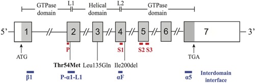
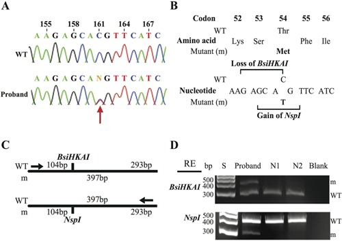
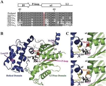
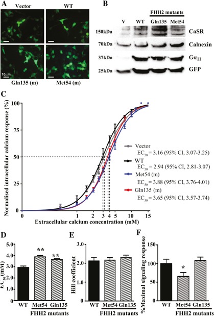
Familial hypocalciuric hypercalcemia (FHH) is a genetically heterogeneous disorder with three variants, FHH1 to FHH3. FHH1 is caused by loss-of-function mutations of the calcium-sensing receptor (CaSR), a G-protein coupled receptor that predominantly signals via G-protein subunit alpha-11 (Gα11 ) to regulate calcium homeostasis. FHH2 is the result of loss-of-function mutations in Gα11 , encoded by GNA11, and to date only two FHH2-associated Gα11 missense mutations (Leu135Gln and Ile200del) have been reported. FHH3 is the result of loss-of-function mutations of the adaptor protein-2 σ-subunit (AP2σ), which plays a pivotal role in clathrin-mediated endocytosis. We describe a 65-year-old woman who had hypercalcemia with normal circulating parathyroid hormone concentrations and hypocalciuria, features consistent with FHH, but she did not have CaSR and AP2σ mutations. Mutational analysis of the GNA11 gene was therefore undertaken, using leucocyte DNA, and this identified a novel heterozygous GNA11 mutation (c.161C>T; p.Thr54Met). The effect of the Gα11 variant was assessed by homology modeling of the related Gαq protein and by measuring the CaSR-mediated intracellular calcium (Ca(2+) i ) responses of HEK293 cells, stably expressing CaSR, to alterations in extracellular calcium (Ca(2+) o ) using flow cytometry. Three-dimensional modeling revealed the Thr54Met mutation to be located at the interface between the Gα11 helical and GTPase domains, and to likely impair GDP binding and interdomain interactions. Expression of wild-type and the mutant Gα11 in HEK293 cells stably expressing CaSR demonstrate that the Ca(2+) i responses after stimulation with Ca(2+) o of the mutant Met54 Gα11 led to a rightward shift of the concentration-response curve with a significantly (p < 0.01) increased mean half-maximal concentration (EC50 ) value of 3.88 mM (95% confidence interval [CI] 3.76-4.01 mM), when compared with the wild-type EC50 of 2.94 mM (95% CI 2.81-3.07 mM) consistent with a loss-of-function. Thus, our studies have identified a third Gα11 mutation (Thr54Met) causing FHH2 and reveal a critical role for the Gα11 interdomain interface in CaSR signaling and Ca(2+) o homeostasis. © 2016 American Society for Bone and Mineral Research.

Keywords: CELL/TISSUE SIGNALING - ENDOCRINE PATHWAYS; DISORDERS OF CALCIUM/PHOSPHATE METABOLISM; PARATHYROID-RELATED DISORDERS; PTH/VIT D/FGF23.


Figures

Figure 1
Figure 1
Schematic representation of the genomic organization of the human GNA11 gene showing the locations of FHH2‐causing mutations. The GNA11 gene consists of 7 exons with the start (ATG) and stop (TGA) codons located in exons 1 and 7, respectively. The GTPase domain (encoded by exon 1, 5' portion of exon 2, 3' portion of exon 4 and exons 5 to 7) is connected to the helical domain (encoded by the 3' portion of exon 2, exon 3, and 5' portion of exon 4) by the linker 1 (L1) and linker 2 (L2) peptides. The locations of the P‐loop (P) (red line), three flexible switch regions (S1 to S3) (red line), and the interdomain interface (comprising portions of the α1, αF, α5, β1, P‐loop, and L1 peptide motifs) (blue line) are shown below the GNA11 exons. The previously reported loss‐of‐function Leu135Gln and Ile200del mutations11 are located in the helical and GTPase domains, respectively, whereas the Thr54Met mutation (bold), identified by this study, is located at the interdomain interface. Coding regions are shaded gray and untranslated regions are represented by open boxes.
Figure 2
Figure 2
Identification of a Thr54Met GNA11 mutation in a FHH proband. (A) DNA sequence analyses revealed a heterozygous C‐to‐T transition at nucleotide c.161 (red arrow) within exon 2 of GNA11. (B) This sequence abnormality was predicted to lead to a missense amino acid substitution of Thr to Met at codon 54, resulting in the loss of a BsiHKAI restriction endonuclease site (GAGCA/C) and gain of an NspI (GCATG/T) restriction endonuclease site. (C) Restriction maps showing that BsiHKAI digestion would result in two products of 104 bp and 293 bp from the wild‐type (WT) sequence but not the mutant (m) sequence. In contrast, NspI digestion results in two products of 104 bp and 293 bp from the m sequence but not the WT sequence. (D) Restriction endonuclease (RE) digest of PCR products of exon 2 of GNA11 demonstrating the heterozygous C‐to‐T transition and confirming its absence in two unrelated unaffected individuals (N1 and N2). S = size marker. For BsiHKAI RE digest the mutant product (397 bp) and WT product (293 bp) are shown; the WT product at 104 bp is not shown. For NspI RE digest the WT product (397 bp) and mutant product (293 bp) are shown; the mutant product at 104 bp is not shown.
Figure 3
Figure 3
Predicted effects of the Thr54Met mutation on the Gα11 protein. (A) Multiple protein sequence alignment of residues contributing to the interdomain interface of Gα‐subunit paralogs. The wild‐type Thr54 (T) and mutant Met54 (M) residues are shown in red. Conserved residues are shaded gray. (B) Overall three‐dimensional structure of the Gα11 protein. The helical (blue) and GTPase (green) domains are connected by the L1 and L2 peptides (gray). The GTPase domain contains the P‐loop (pink), which binds GDP (black) at the interdomain interface (comprising parts of α1, α5, αF, P‐loop, L1, and β1 peptide motifs). The reported Leu135Gln and Ile200del mutations11 are shown in purple, and the Thr54Met mutation is shown in red. (C) Close‐up views of the Gα11 interdomain interface, which show the wild‐type Thr54 residue, located in the GTPase domain, form polar contacts (hatched black lines) with GDP and the Arg181 residue (yellow) of the helical domain αF helix. Substitution of the wild‐type Thr54 residue with the mutant Met54 residue (orange) is predicted to disrupt these polar contacts, thereby impairing interdomain interactions and the binding of GDP.
Figure 4
Figure 4
Functional characterization of wild‐type and FHH2‐associated mutant Gα11 proteins. (A) Fluorescence microscopy of HEK293 cells stably expressing CaSR (HEK293‐CaSR) and transiently transfected with wild‐type (WT) or FHH2‐associated mutant (Met54 and Gln135) pBI‐CMV2‐GNA11‐GFP constructs, or with vector only. GFP expression in these cells indicates successful transfection and expression by these constructs. Scale bar = 10 µm. (B) Western blot analyses of whole‐cell lysates using antibodies to CaSR, calnexin, Gα11, and GFP. Transient transfection of WT or FHH2‐associated mutant constructs resulted in overexpression of Gα11 when normalized to calnexin expression. (C) Concentration‐response curves showing normalized Ca2+ i response to changes in [Ca2+]o of HEK293‐CaSR cells transfected with WT or FHH2‐associated Gα11 mutants. The Ca2+ i responses are shown as the mean ± SEM of 4 independent transfections. The FHH2‐associated Gα11 mutants (Met54 and Gln135) led to a rightward shift of the concentration‐response curves (blue and red, respectively) when compared with WT Gα11 (black), which harbors Thr and Leu residues at codons 54 and 135, respectively. (D) The Met54 and Gln135 mutants (shaded bars) were associated with significantly increased EC50 values compared with cells expressing WT Gα11 (black bar). (E) The Hill coefficients of the wild‐type and mutant Gα11 proteins were similar (ie, not significantly different). (F) The Met54 mutant was associated with a significantly reduced maximal signaling response compared with WT and mutant Gln135 Gα11 proteins. *p < 0.05, **p < 0.01.

References

    1. Hannan FM, Thakker RV. Calcium‐sensing receptor (CaSR) mutations and disorders of calcium, electrolyte and water metabolism. Best Pract Res Clin Endocrinol Metab. 2013; 27(3):359–71. - PubMed
    1. Eastell R, Brandi ML, Costa AG, D'Amour P, Shoback DM, Thakker RV. Diagnosis of asymptomatic primary hyperparathyroidism: proceedings of the Fourth International Workshop. J Clin Endocrinol Metab. 2014; 99(10):3570–9. - PubMed
    1. Hannan FM, Nesbit MA, Zhang C, et al. Identification of 70 calcium‐sensing receptor mutations in hyper‐ and hypo‐calcaemic patients: evidence for clustering of extracellular domain mutations at calcium‐binding sites. Hum Mol Genet. 2012; 21(12):2768–78. - PubMed
    1. Wu DQ, Lee CH, Rhee SG, Simon MI. Activation of phospholipase C by the alpha subunits of the Gq and G11 proteins in transfected Cos‐7 cells. J Biol Chem. 1992; 267(3):1811–7. - PubMed
    1. Hofer AM, Brown EM. Extracellular calcium sensing and signalling. Nat Rev Mol Cell Biol. 2003; 4(7):530–8. - PubMed

Publication types

Supplementary concepts