. 2016 Apr;15(4):1232-45.
doi: 10.1074/mcp.M115.055954. Epub 2016 Jan 4.

Proteome-wide Identification of Novel Ceramide-binding Proteins by Yeast Surface cDNA Display and Deep Sequencing

Affiliations

Proteome-wide Identification of Novel Ceramide-binding Proteins by Yeast Surface cDNA Display and Deep Sequencing

Scott Bidlingmaier et al. Mol Cell Proteomics. 2016 Apr.

Abstract

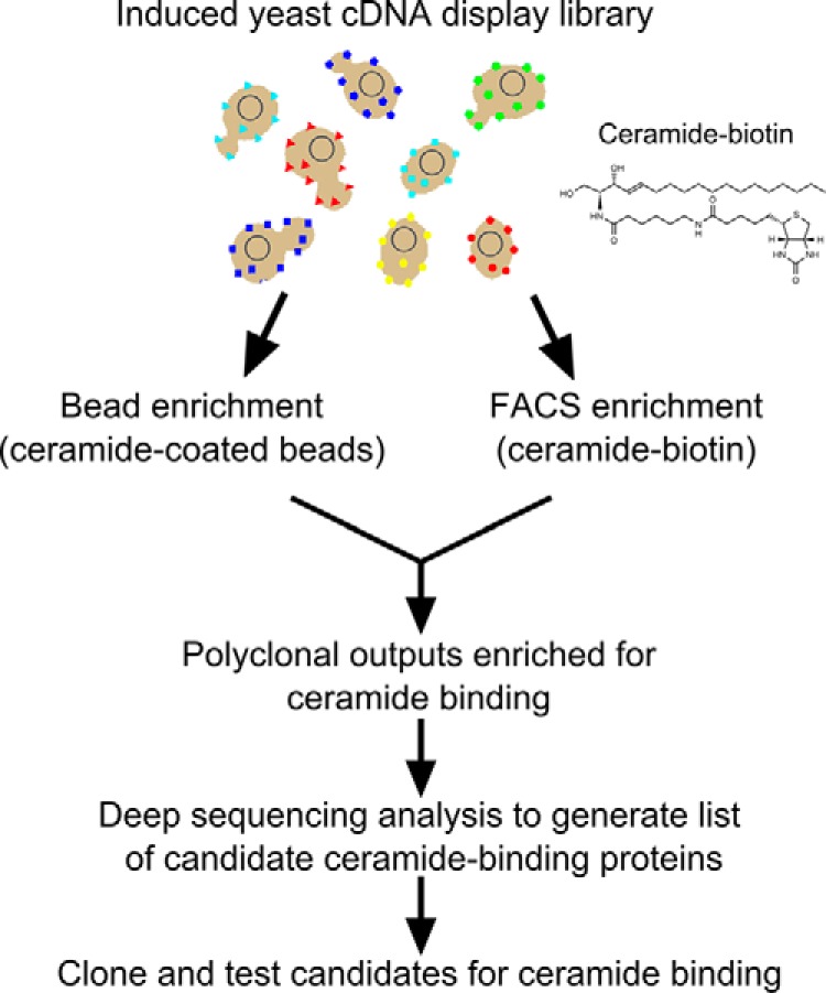
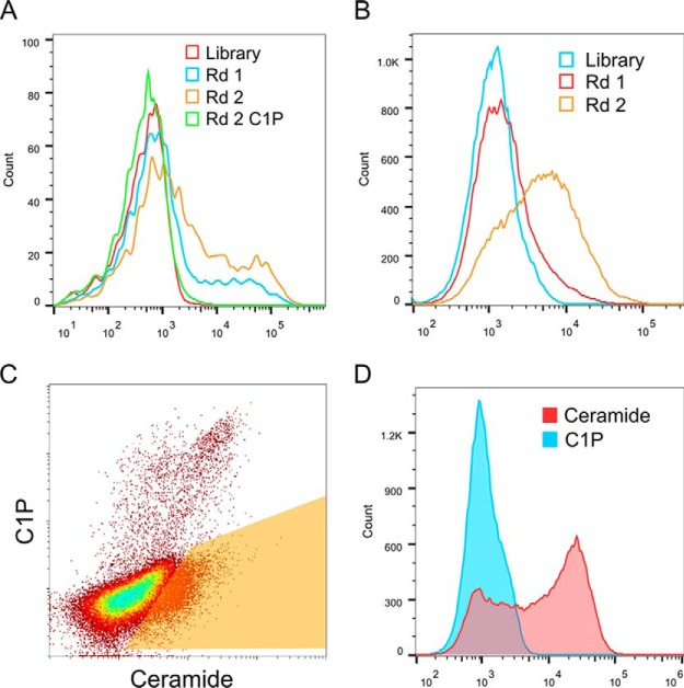
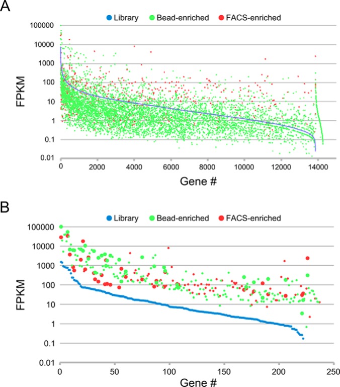
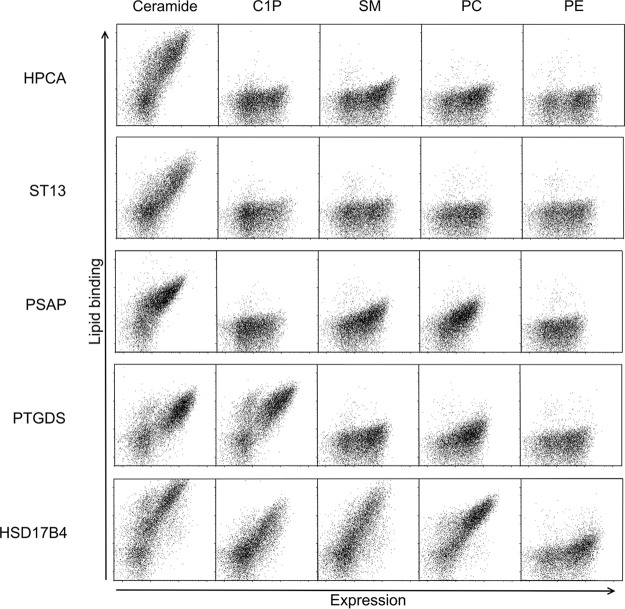
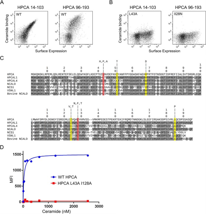
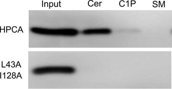
Although the bioactive sphingolipid ceramide is an important cell signaling molecule, relatively few direct ceramide-interacting proteins are known. We used an approach combining yeast surface cDNA display and deep sequencing technology to identify novel proteins binding directly to ceramide. We identified 234 candidate ceramide-binding protein fragments and validated binding for 20. Most (17) bound selectively to ceramide, although a few (3) bound to other lipids as well. Several novel ceramide-binding domains were discovered, including the EF-hand calcium-binding motif, the heat shock chaperonin-binding motif STI1, the SCP2 sterol-binding domain, and the tetratricopeptide repeat region motif. Interestingly, four of the verified ceramide-binding proteins (HPCA, HPCAL1, NCS1, and VSNL1) and an additional three candidate ceramide-binding proteins (NCALD, HPCAL4, and KCNIP3) belong to the neuronal calcium sensor family of EF hand-containing proteins. We used mutagenesis to map the ceramide-binding site in HPCA and to create a mutant HPCA that does not bind to ceramide. We demonstrated selective binding to ceramide by mammalian cell-produced wild type but not mutant HPCA. Intriguingly, we also identified a fragment from prostaglandin D2synthase that binds preferentially to ceramide 1-phosphate. The wide variety of proteins and domains capable of binding to ceramide suggests that many of the signaling functions of ceramide may be regulated by direct binding to these proteins. Based on the deep sequencing data, we estimate that our yeast surface cDNA display library covers ∼60% of the human proteome and our selection/deep sequencing protocol can identify target-interacting protein fragments that are present at extremely low frequency in the starting library. Thus, the yeast surface cDNA display/deep sequencing approach is a rapid, comprehensive, and flexible method for the analysis of protein-ligand interactions, particularly for the study of non-protein ligands.


Conflict of interest statement

The authors declare that they have no conflicts of interest with the contents of this article.

Figures

Fig. 1.
Fig. 1.
Flow chart of strategy for identification of ceramide-binding protein fragments.
Fig. 2.
Fig. 2.
Generation of polyclonal yeast display selection outputs enriched for ceramide binding. A, FACS analysis of ceramide-coated bead enrichment outputs. The starting library and polyclonal selection outputs from the first and second rounds were tested for binding to 200 nm ceramide by FACS. The second round output was also tested for binding to 200 nm C1P. B, FACS analysis of FACS-sorted enrichment outputs. The starting library and polyclonal selection outputs from the first and second rounds were tested for binding to 200 nm ceramide by FACS. C, two-color competitive selection with both ceramide and C1P. The FACS-enriched second round polyclonal output was incubated with 200 nm labeled ceramide and C1P, and the ceramide-specific binding population was gated and sorted. D, FACS analysis of the polyclonal competitive selection output. The polyclonal output of the third round competitive selection was tested by FACS using 200 nm ceramide and C1P.
Fig. 3.
Fig. 3.
Graphical representations of starting library, bead-enriched, and FACS-enriched gene-level FPKM values calculated from deep sequencing data sets. The FPKM data for the individual genes is arranged by starting library FPKM (high to low from left to right). A, FPKM values for the 14,267 unique non-zero genes from the combined starting library, bead-enriched, and FACS-enriched analyzed data sets. B, FPKM values of the 234 unique genes that meet frequency and enrichment cutoffs and whose recovered nucleotide sequences encode protein fragments derived from the corresponding genes. Larger data points indicate candidates that meet the frequency and enrichment cutoffs in both the bead-enriched and FACS-enriched data sets.
Fig. 4.
Fig. 4.
FACS plots depicting binding of ceramide, C1P, SM, phosphatidylcholine (PC), and PE by selected candidate ceramide-binding protein fragments displayed on yeast. All the lipids were tested at a concentration of 200 nm. Lipid binding is represented in the y axis, and surface expression level (Xpress epitope) is represented in the x axis.
Fig. 5.
Fig. 5.
Isolation of non-ceramide binding HPCA mutants. A, binding of HPCA 1 + 2 (amino acids 14–103) and HPCA 3 + 4 (amino acids 96–193) to 200 nm ceramide. Ceramide binding is represented by the y axis, and surface expression (Xpress epitope) is represented by the x axis. B, binding of HPCA 1 + 2 L43A and HPCA 3 + 4 I128N to 200 nm ceramide. C, alignment of candidate and verified ceramide-binding NCS family proteins showing the position of mutations in HPCA that reduce ceramide binding. Amino acids identical to HPCA are shaded gray. Mutations in amino acid residues shaded yellow or red dramatically reduce ceramide binding in the context of HPCA 1 + 2 or HPCA 3 + 4. Amino acid residues shaded red were additionally tested in combination in full-length HPCA. One-letter amino acid codes above each shaded mutation indicate the amino acid substitutions recovered that significantly reduce ceramide binding. D, measurement of ceramide affinity for yeast-displayed full-length HPCA and HPCA L43A/I128A mutant.
Fig. 6.
Fig. 6.
Pulldown of epitope-tagged HPCA and HPCA L43A/I128A mutant from transfected HeLa extracts. Beads used for capture were coated with ceramide (Cer), C1P, and SM. Extract equivalent to the total amount included in the pulldown experiments is shown in the 1st lane.

References

    1. Galadari S., Rahman A., Pallichankandy S., and Thayyullathil F. (2015) Tumor suppressive functions of ceramide: evidence and mechanisms. Apoptosis 20, 689–711 - PubMed
    1. Morad S. A., and Cabot M. C. (2013) Ceramide-orchestrated signalling in cancer cells. Nat. Rev. Cancer 13, 51–65 - PubMed
    1. Saddoughi S. A., and Ogretmen B. (2013) Diverse functions of ceramide in cancer cell death and proliferation. Adv. Cancer Res. 117, 37–58 - PubMed
    1. Maceyka M., and Spiegel S. (2014) Sphingolipid metabolites in inflammatory disease. Nature 510, 58–67 - PMC - PubMed
    1. Mencarelli C., and Martinez-Martinez P. (2013) Ceramide function in the brain: when a slight tilt is enough. Cell. Mol. Life Sci. 70, 181–203 - PMC - PubMed

Publication types