. 2016 Feb 9;113(6):1642-7.
doi: 10.1073/pnas.1516979113. Epub 2016 Jan 4.

Respiratory syncytial virus infection enhances Pseudomonas aeruginosa biofilm growth through dysregulation of nutritional immunity

Affiliations

Respiratory syncytial virus infection enhances Pseudomonas aeruginosa biofilm growth through dysregulation of nutritional immunity

Matthew R Hendricks et al. Proc Natl Acad Sci U S A. .

Abstract

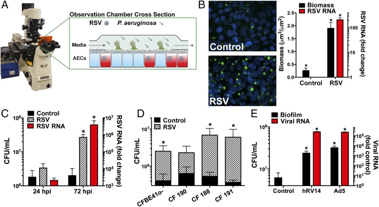
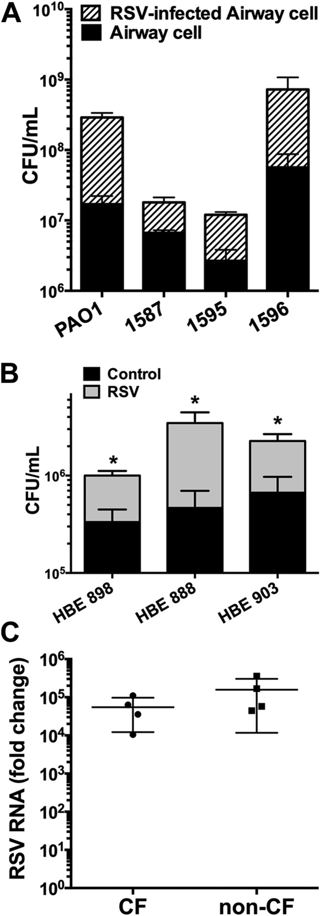
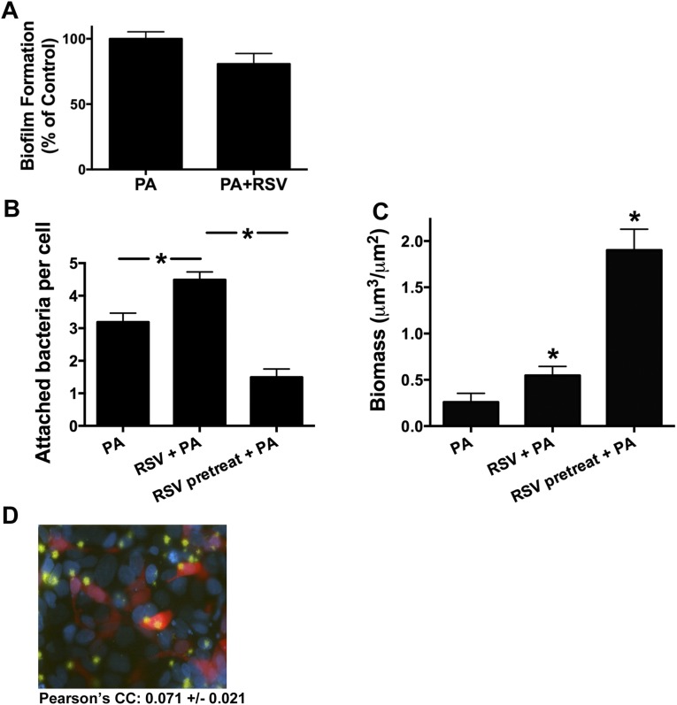
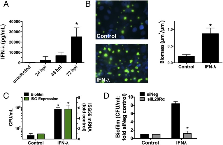
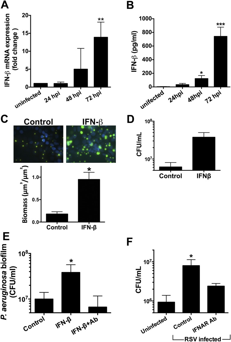
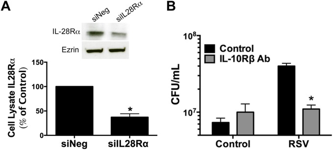
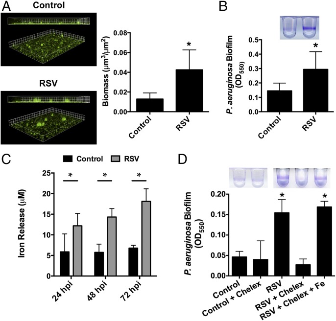
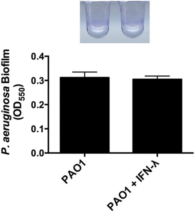
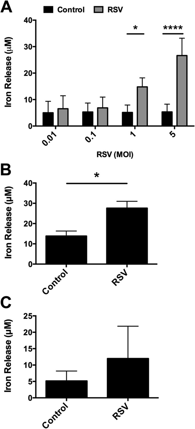
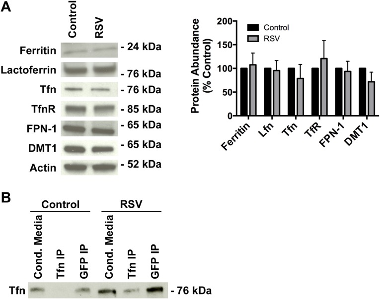
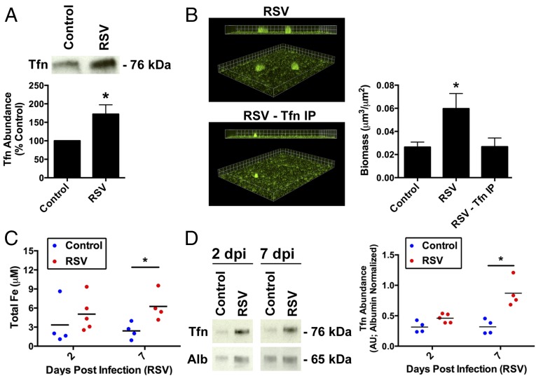
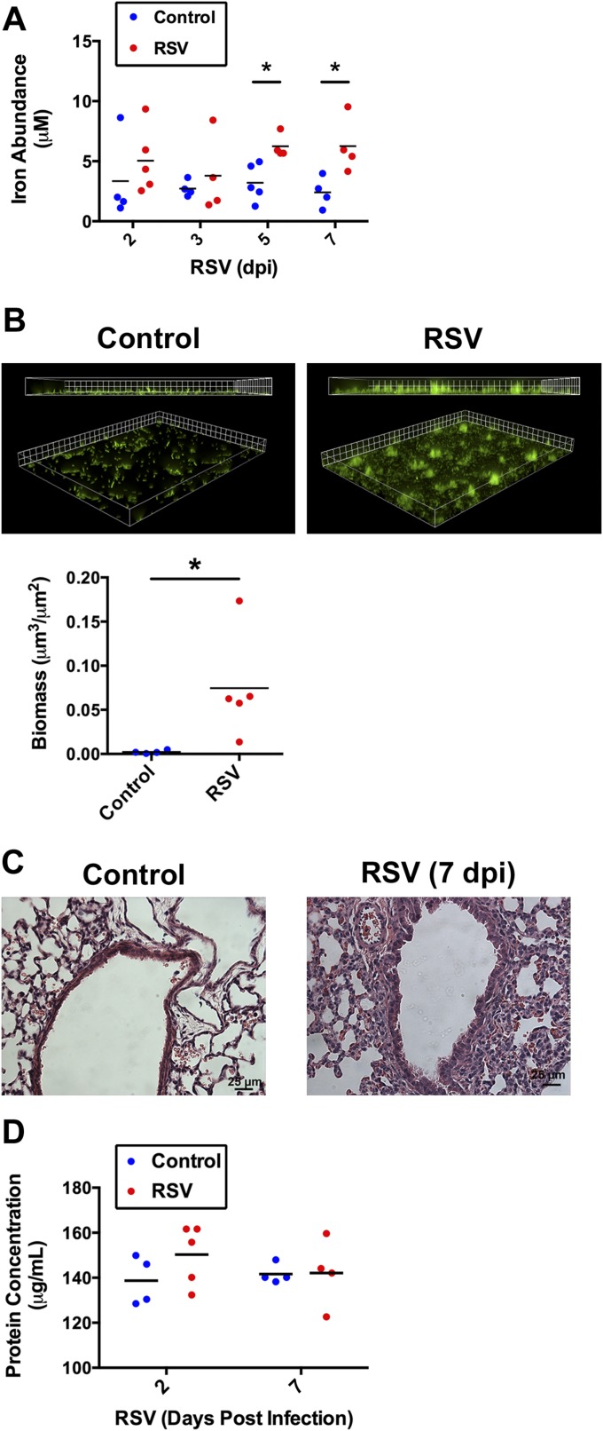
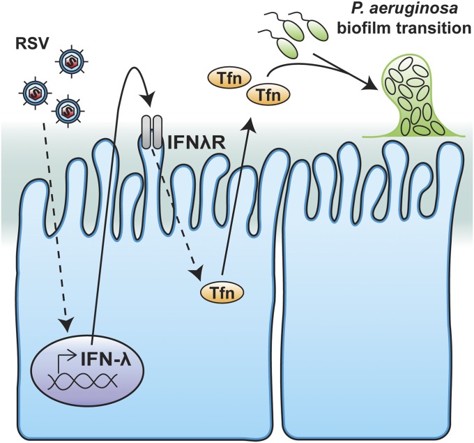
Clinical observations link respiratory virus infection and Pseudomonas aeruginosa colonization in chronic lung disease, including cystic fibrosis (CF) and chronic obstructive pulmonary disease. The development of P. aeruginosa into highly antibiotic-resistant biofilm communities promotes airway colonization and accounts for disease progression in patients. Although clinical studies show a strong correlation between CF patients' acquisition of chronic P. aeruginosa infections and respiratory virus infection, little is known about the mechanism by which chronic P. aeruginosa infections are initiated in the host. Using a coculture model to study the formation of bacterial biofilm formation associated with the airway epithelium, we show that respiratory viral infections and the induction of antiviral interferons promote robust secondary P. aeruginosa biofilm formation. We report that the induction of antiviral IFN signaling in response to respiratory syncytial virus (RSV) infection induces bacterial biofilm formation through a mechanism of dysregulated iron homeostasis of the airway epithelium. Moreover, increased apical release of the host iron-binding protein transferrin during RSV infection promotes P. aeruginosa biofilm development in vitro and in vivo. Thus, nutritional immunity pathways that are disrupted during respiratory viral infection create an environment that favors secondary bacterial infection and may provide previously unidentified targets to combat bacterial biofilm formation.

Keywords: Pseudomonas aeruginosa; biofilm; cystic fibrosis; nutritional immunity; respiratory syncytial virus.


Conflict of interest statement

The authors declare no conflict of interest.

Figures

Fig. 1.
Fig. 1.
Respiratory viral infection promotes the growth of P. aeruginosa biofilm on AECs. (A) The setup for live-cell biotic biofilm imaging and a cross-sectional view of the micro-observation chamber. (B, Right) P. aeruginosa (GFP) biofilms imaged by live-cell microscopy after 6 h of growth. AEC cell nuclei are shown with Hoechst (blue) staining. (Left) Biofilm biomass was quantified using COMSTAT (black bars, left y axis). RSV RNA was measured by quantitative RT-PCR (qRT-PCR) to assess RSV infection (red bar, right y axis). (C) RSV-enhanced biofilm growth is time dependent. In a static coculture biofilm assay, AECs were infected with RSV (striped bars) or were mock-infected [Eagle's minimal essential media (MEM) control; black bars] for the indicated times followed by P. aeruginosa infection. P. aeruginosa biofilm was assessed by cfu enumeration (Left y axis). RSV RNA was measured by qRT-PCR to assess RSV infection (red bars, right y axis). (D) RSV stimulates the growth of P. aeruginosa biofilm on well-differentiated CF HBEs. P. aeruginosa biofilms were grown on primary CF HBEs using the static coculture biofilm assay and were quantified by cfu enumeration. (E) Other respiratory viruses enhance the growth of P. aeruginosa biofilm on AECs. P. aeruginosa biofilms were grown in a static coculture biofilm assay on AECs infected with hRV or AdV or were mock-infected (MEM control). Biofilms were quantified by cfu enumeration (black bars, left y axis). Viral RNA was measured by qRT-PCR to assess virus infection (red bars, right y axis). RSV, RSV-infected AECs. For all experiments n ≥ 3. Data are presented as mean ± SD; *P < 0.05 versus control.
Fig. S1.
Fig. S1.
RSV-enhanced growth of P. aeruginosa biofilm is seen in CF clinical isolates and is dose-dependent. (A) RSV infection promotes biofilm formation by CF P. aeruginosa clinical isolates in a static coculture biofilm assay. In a static coculture biofilm assay, AECs were infected with RSV (striped bars) or were mock-infected (MEM control; black bars) for 72 h and then were infected with the indicated clinical isolates of P. aeruginosa. (B) RSV enhances the growth of P. aeruginosa biofilm on well-differentiated non-CF HBEs. P. aeruginosa biofilms were grown on non-CF HBEs in a static coculture biofilm assay following 72-h RSV infection and were quantified by cfu enumeration. (C) RSV RNA was measured by qRT-PCR to assess RSV infection, which is equivalent in CF and non-CF HBE cells. RSV, RSV-infected AECs. n ≥ 3 for all experiments. Data are presented as mean ± SD; *P < 0.05.
Fig. S2.
Fig. S2.
RSV-stimulated biofilm growth is not caused by direct viral–P. aeruginosa interaction. (A) P. aeruginosa biofilms were grown in MEM ± RSV for 24 h in the 96-well microtiter biofilm assay. Biofilms were quantified by crystal violet staining and were measured as absorbance at 550 nm. Biofilm growth is presented as the percentage of growth in MEM alone. (B) P. aeruginosa attachment is reduced on AECs with preceding RSV infection. AECs were infected with P. aeruginosa (PA), simultaneously with RSV and P. aeruginosa (RSV + PA), or with RSV before infection with P. aeruginosa (RSV pretreat + PA). P. aeruginosa (GFP) adherence on AECs was imaged by live-cell microscopy after 1 h of bacterial infection. Bacterial counts were normalized to the number of epithelial cells per field by counting nuclei (10 fields were counted per treatment, repeated three times). (C) The growth of P. aeruginosa biofilm is enhanced only on AECs with preceding RSV infection. P. aeruginosa biofilms were imaged by live-cell microscopy after 6 h of growth on AECs, and biofilm biomass was quantified by COMSTAT. AECs were infected and are labeled along the x axis as in B. (D) P. aeruginosa biofilms are randomly distributed on AECs infected with RSV. Cells were infected with RSV-RFP for 24 h followed by PAO1-GFP infection, and biofilm growth (green) was imaged after 5 h by live-cell microcopy. Hoechst (blue) staining shows epithelial cell nuclei. For all experiments n ≥ 3. Data are presented as mean ± SD; *P < 0.05.
Fig. S3.
Fig. S3.
The AEC monolayer is intact during RSV infection. (A) Epithelial integrity was assessed by the lactate dehydrogenase release assay at 72 hpi for various doses of RSV (MOI = 0.001–5). Means are not significantly different from the uninfected control. (B) Transepithelial electrical resistance measurements were used to assess epithelial integrity over the course of 72-h RSV infection. AECs were infected with RSV (red triangles) or were mock-infected (MEM control; black circles) for the indicated times.
Fig. 2.
Fig. 2.
Type III IFN (IFN-λ) signaling stimulates the growth of P. aeruginosa biofilm. (A) RSV infection induces IFN-λ secretion from AECs. Cells were infected with RSV for the indicated number of hours (hpi), and IFN-λ1/3 (IL-29/28B) release was measured by ELISA. (B and C) IFN-λ treatment stimulates the growth of P. aeruginosa biofilm on AECs. P. aeruginosa biofilm growth increased on AECs treated for 12 h with IFN-λ1 (100 ng/mL), as assessed by live-cell microscopy (B) or a static coculture biofilm assay (C). Epithelial cell nuclei are shown with Hoechst (blue) staining. The P. aeruginosa biofilm (GFP, green) biomass was calculated for each condition using COMSTAT (B). P. aeruginosa biofilm was assessed by cfu enumeration (black bars, left y axis) (C). IFN-λ1 signaling was confirmed with ISG56 induction by qRT-PCR (green bars, right y axis) (C). (D) Signaling via IL-28Rα is required for biofilm growth during IFN-λ treatment. AECs were transfected with scrambled siRNA (siNeg) or siRNA targeting IL-28Rα (siIL28Rα) and were treated with IFN-λ1 (100 ng/mL) for 12 h, and P. aeruginosa biofilms were grown in a static coculture biofilm assay. P. aeruginosa biofilm growth was quantified by cfu enumeration and displayed as fold change compared with siNeg-transfected cells. IFN-λ, IFN-λ–treated AECs. For all experiments n ≥ 3. Data are presented as mean ± SD; *P < 0.05.
Fig. S4.
Fig. S4.
IFN-β treatment enhances the growth of P. aeruginosa biofilm. (A and B) RSV infection induces IFN-β expression and secretion from AECs. AECs were infected with RSV for the indicated number of hours. (A) IFN-β expression was measured by qPCR. (B) Protein release was measured by ELISA. (C and D) The growth of P. aeruginosa biofilm increased on AECs treated with IFN-β (1,000 U/mL) for 18 h, as measured by live-cell microscopy (C) or in the static coculture biofilm assay (D). (E) Signaling through IFNAR is required for biofilm growth induced by IFN-β treatment. AECs were treated with IFNAR-neutralizing antibody (5 μg/mL) during 18-h treatment with IFN-β (1,000 U/mL), and P. aeruginosa biofilms were grown in a static coculture biofilm assay. Control corresponds to mock IFN-β treatment in the absence of IFNAR-neutralizing antibody. The growth of P. aeruginosa biofilm was quantified by cfu enumeration. (F) Neutralizing IFNAR prevents the RSV-stimulated growth of P. aeruginosa biofilm. AECs were infected with RSV (RSV infected) and were treated with neutralizing IFNAR-neutralizing antibody (IFNAR Ab; 5 μg/mL); P. aeruginosa biofilms were grown in a static coculture biofilm assay. The growth of P. aeruginosa biofilm was quantified by cfu enumeration. Uninfected, mock infection (MEM control). n ≥ 3 for all experiments. Data are presented as mean ± SD; *P < 0.05.
Fig. S5.
Fig. S5.
IFN-λ signaling is required for the growth of virus-stimulated P. aeruginosa biofilm. Signaling through IL-28Rα is required for biofilm growth during IFN-λ treatment. AECs were transfected with scrambled siRNA (siNeg) or siRNA targeting IL-28Rα (siIL28Rα). (A) siRNA-mediated knockdown of IL-28Rα was assessed by measuring protein abundance for each condition by Western blot and is displayed as the percent of IL-28Rα in cells transfected with siNeg (control). Biofilm formation in these conditions was assayed in Fig. 3D. (B) Neutralizing IFN-λR prevents RSV-enhanced formation of P. aeruginosa biofilm. Cells were infected with RSV or were mock-infected (MEM control) for 72 h and were treated with IL-10Rβ–neutralizing antibody (IL-10Rβ Ab; 10 μg/mL) (gray bars) or were left untreated (black bars); P. aeruginosa biofilms were grown in static coculture biofilm assays. The growth of P. aeruginosa biofilm was quantified by cfu enumeration. RSV, RSV-infected AECs. For all experiments n ≥ 3. Data are presented as mean ± SD; *P < 0.05.
Fig. 3.
Fig. 3.
RSV infection enhances iron release from AECs to stimulate biofilm growth. (A and B) RSV infection stimulates the release of a biofilm-stimulatory factor that promotes P. aeruginosa formation. AECs were infected with RSV or were mock-infected (MEM control) for 72 h, and the apical CM was collected. (A) P. aeruginosa (GFP) was grown in the presence of CM in static abiotic biofilm assays. Epifluorescence microscopy was used to measure the growth of P. aeruginosa biofilm (GFP, green), and biomass was quantified using Nikon Elements (grid unit = 9 μm). (B) P. aeruginosa biofilms were grown in CM for 24 h in a 96-well microtiter biofilm assay. (C) Total iron was increased in apical CM collected from AECs infected with RSV or mock-infected (MEM control) for the indicated number of hours postinfection. (D) Iron in RSV CM is required for the growth of P. aeruginosa biofilm. Ninety-six–well microtiter biofilm assays were performed to measure the growth of P. aeruginosa in CM. Divalent metal cations were chelated with Chelex-100 (labeled “Chelex” in the figure), and iron was added back with FeCl3 (8 μM) after Chelex-100 treatment. RSV, RSV-infected AECs. For all experiments n ≥ 3. Data are presented as mean ± SD; *P < 0.05.
Fig. S6.
Fig. S6.
IFN-λ does not interact directly with P. aeruginosa and enhance biofilm growth. P. aeruginosa biofilms were grown in the presence or absence of IFN-λ (200 ng/mL) diluted in MEM for 24 h in the 96-well microtiter biofilm assay. Biofilm growth was measured as absorbance at 550 nm following crystal violet staining and is presented as OD550. n = 3; data are presented as mean ± SD.
Fig. S7.
Fig. S7.
RSV infection enhances iron release from primary CF and non-CF HBEs. (A) Total iron released in the apical CM collected from AECs infected with RSV at the indicated MOI or mock-infected (MEM control). (B and C) Total iron in the apical CM collected from primary CF (B) and non-CF (C) HBEs infected with RSV or mock-infected (MEM control). n = 3; data are presented as mean ± SD; *P < 0.05.
Fig. S8.
Fig. S8.
RSV infection disrupts iron homeostasis in AECs. (A) RSV infection does not affect the abundance of iron transporter proteins in AECs infected with RSV, as measured by Western blot analysis. n = 3; data are presented as mean ± SD. (B) Transferrin depletion reduces the growth of P. aeruginosa biofilm in RSV CM. Transferrin depletion was achieved by immunoprecipitation of transferrin from CM using an anti-transferrin polyclonal antibody. Western blot analysis was used verify transferrin depletion from CM. Corresponding biofilm growth is shown in Fig. 4B.
Fig. 4.
Fig. 4.
Transferrin release increases in response to virus infection in vitro and in vivo. (A and B) AECs were infected with RSV or were mock-infected (MEM control) for 72 h and then apical CM was collected. (A) RSV infection increases transferrin abundance in apical CM, as measured by Western blot analysis. (B) Transferrin depletion reduces the growth of P. aeruginosa biofilm in RSV CM. Apical CM was collected from RSV-infected AECs depleted of transferrin by immunoprecipitation (RSV-Tfn IP). P. aeruginosa (GFP) biofilms were grown in transferrin-replete and -depleted RSV CM in static abiotic biofilm assays. Biofilm biomass was quantified using Nikon Elements (grid unit = 8.5 μm). (C and D) Total iron (C) and transferrin abundance (D) were increased in BALF recovered from neonatal mice infected with RSV or mock-infected (PBS control) for the indicated number of days postinfection (dpi), as measured by iron assay or Western blot analysis, respectively. Horizontal lines indicate mean values. Alb, albumin; RSV, RSV infection; Tfn, transferrin. All experiments were repeated, with at least four mice per group; *P < 0.05.
Fig. S9.
Fig. S9.
BALF recovered from neonatal mice infected with RSV stimulates the growth of P. aeruginosa biofilm. (A) Iron release over the time course of RSV infection in vivo. Mice were infected with RSV for 7 d, and at 2–7 dpi BALF was collected and assayed for iron content. n = 4 or 5 mice per treatment group. (B) P. aeruginosa (GFP) biofilms grown in BALF recovered from neonatal mice infected with RSV 5 dpi in static abiotic biofilm assays. Biofilm biomass was quantified using Nikon Elements (grid unit = 7.5 μm). (C) Lung pathology in mice following RSV infection. Mice were infected with RSV for 7 d; then lungs were harvested, sliced, and stained with H&E. Each panel represents an individual mouse from the indicated group. (Scale bar, 25 μm.) (D) At 2 and 7 dpi BALF was collected and assayed for protein content by Bradford assay. n = 4 or 5 mice per treatment group. Means are not significantly different from PBS controls. *P < 0.05.
Fig. S10.
Fig. S10.
Proposed model for RSV-induced growth of P. aeruginosa biofilm in the lung. Respiratory virus infection and the subsequent induction of IFN signaling pathways results in the increased formation of bacterial biofilm by a mechanism of dysregulated host nutritional immunity mechanisms, leading to increased release of iron-bound transferrin during virus infection. The release of transferrin promotes the transition of P. aeruginosa to a biofilm mode of growth.

Comment in

Similar articles

Cited by

References

    1. Tang AC, et al. Current concepts: Host-pathogen interactions in cystic fibrosis airways disease. Eur Respir Rev. 2014;23(133):320–332. - PMC - PubMed
    1. Suárez-Arrabal MC, et al. Nasopharyngeal bacterial burden and antibiotics: Influence on inflammatory markers and disease severity in infants with respiratory syncytial virus bronchiolitis. J Infect. 2015;71(4):458–469. - PubMed
    1. Varkey JB, Varkey B. Viral infections in patients with chronic obstructive pulmonary disease. Curr Opin Pulm Med. 2008;14(2):89–94. - PubMed
    1. van Ewijk BE, van der Zalm MM, Wolfs TF, van der Ent CK. Viral respiratory infections in cystic fibrosis. J Cyst Fibros. 2005;4(Suppl 2):31–36. - PMC - PubMed
    1. Wat D, et al. The role of respiratory viruses in cystic fibrosis. J Cyst Fibros. 2008;7(4):320–328. - PMC - PubMed

Publication types

MeSH terms

LinkOut - more resources