. 2016 Jan 19;113(3):E348-57.
doi: 10.1073/pnas.1523321113. Epub 2016 Jan 4.

Separable roles for Mycobacterium tuberculosis ESX-3 effectors in iron acquisition and virulence

Affiliations

Separable roles for Mycobacterium tuberculosis ESX-3 effectors in iron acquisition and virulence

JoAnn M Tufariello et al. Proc Natl Acad Sci U S A. .

Abstract

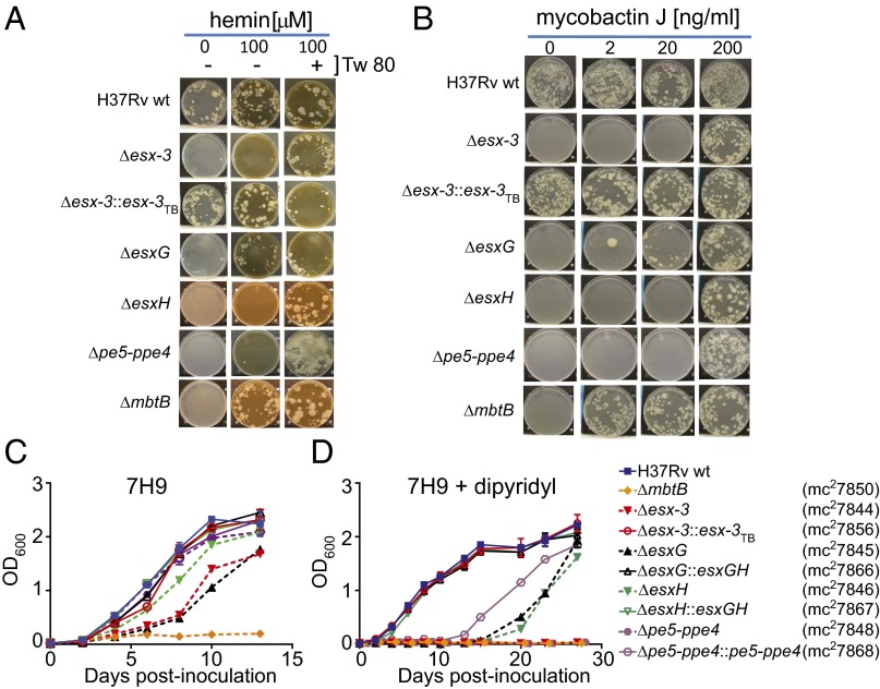
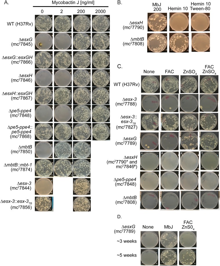
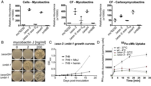
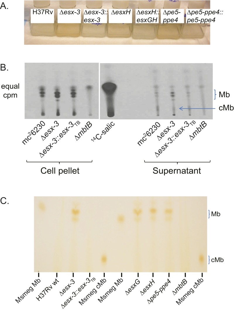
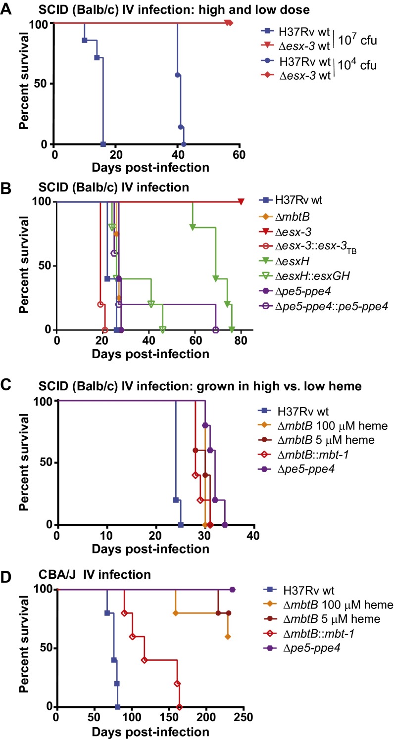
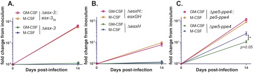
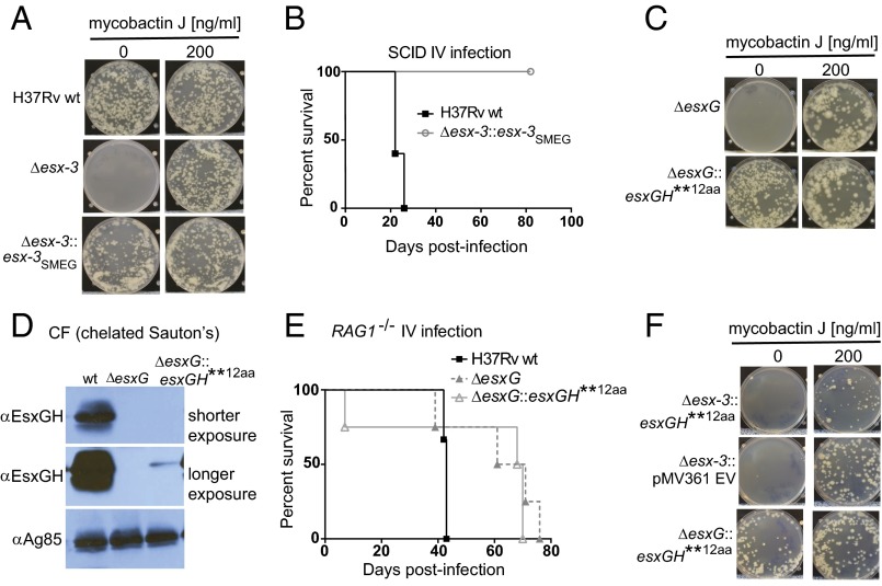
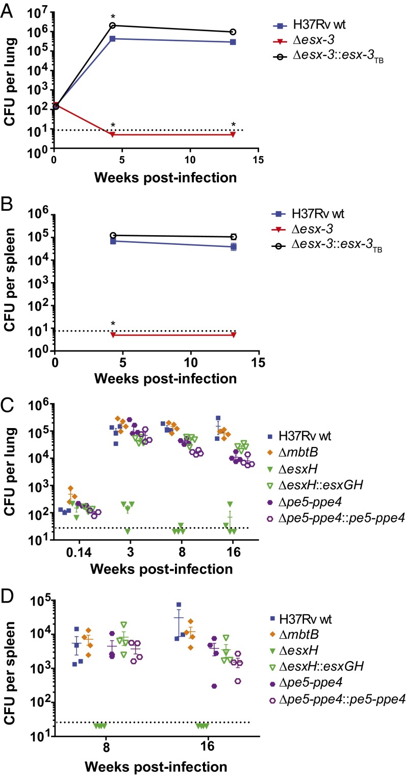
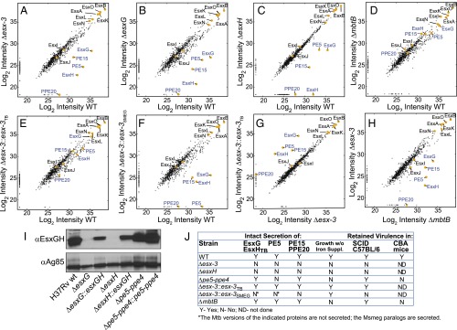
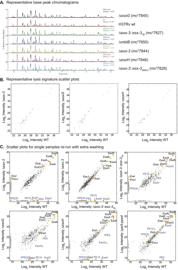
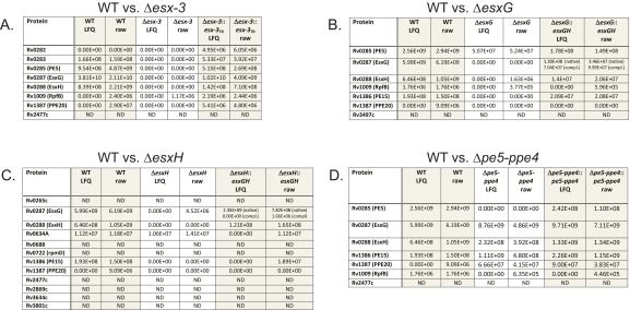
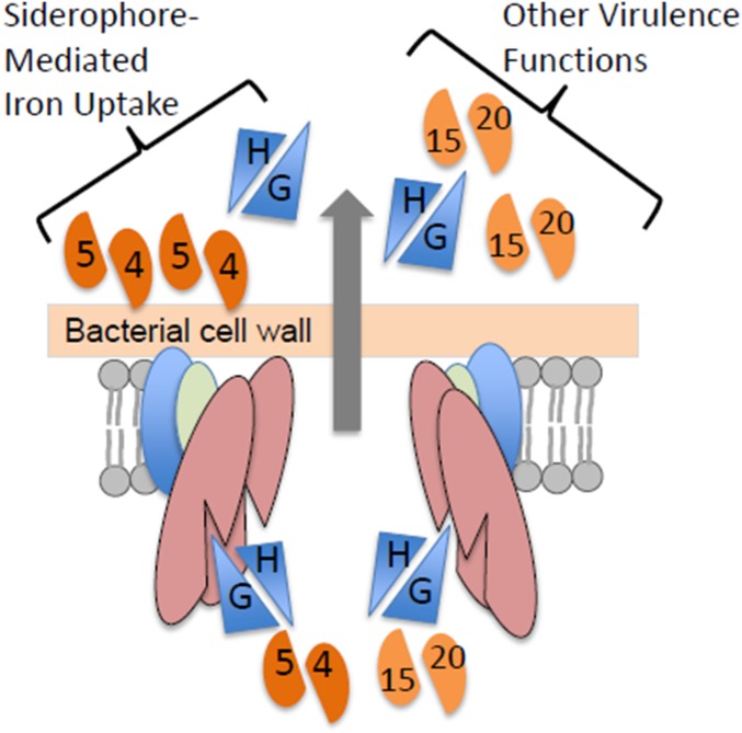
Mycobacterium tuberculosis (Mtb) encodes five type VII secretion systems (T7SS), designated ESX-1-ESX-5, that are critical for growth and pathogenesis. The best characterized is ESX-1, which profoundly impacts host cell interactions. In contrast, the ESX-3 T7SS is implicated in metal homeostasis, but efforts to define its function have been limited by an inability to recover deletion mutants. We overcame this impediment using medium supplemented with various iron complexes to recover mutants with deletions encompassing select genes within esx-3 or the entire operon. The esx-3 mutants were defective in uptake of siderophore-bound iron and dramatically accumulated cell-associated mycobactin siderophores. Proteomic analyses of culture filtrate revealed that secretion of EsxG and EsxH was codependent and that EsxG-EsxH also facilitated secretion of several members of the proline-glutamic acid (PE) and proline-proline-glutamic acid (PPE) protein families (named for conserved PE and PPE N-terminal motifs). Substrates that depended on EsxG-EsxH for secretion included PE5, encoded within the esx-3 locus, and the evolutionarily related PE15-PPE20 encoded outside the esx-3 locus. In vivo characterization of the mutants unexpectedly showed that the ESX-3 secretion system plays both iron-dependent and -independent roles in Mtb pathogenesis. PE5-PPE4 was found to be critical for the siderophore-mediated iron-acquisition functions of ESX-3. The importance of this iron-acquisition function was dependent upon host genotype, suggesting a role for ESX-3 secretion in counteracting host defense mechanisms that restrict iron availability. Further, we demonstrate that the ESX-3 T7SS secretes certain effectors that are important for iron uptake while additional secreted effectors modulate virulence in an iron-independent fashion.

Keywords: ESX-3; Mycobacterium tuberculosis; mycobactin; siderophore; type VII secretion system.


Conflict of interest statement

The authors declare no conflict of interest.

Figures

Fig. 1.
Fig. 1.
Defined iron sources rescue the essentiality of esx-3 region mutants. (A and B) The indicated Mtb strains H37Rv WT, Δesx-3 (mc27788), Δesx-3::esx-3TB (mc27827), ΔesxG (mc27789), ΔesxH (mc27790), Δpe5ppe4 (mc27792), and ΔmbtB (mc27808) (Fig. S1 and Table S1) were plated on 7H10 medium with or without 100 μM hemin and Tween 80 (0.05%) (Tw 80) (A) or with increasing concentrations of MbJ (B). Some late-appearing contamination was present when Δpe5ppe4 was plated on medium containing hemin and Tween 80 in A. (C and D) The indicated strains, which differ from those used in the plating experiments shown in A and B in that the sacB-hygR cassettes were removed, were grown in 7H9 medium (C) or 7H9 medium supplemented with 100 μM 2,2′-dipyridyl (D). Data points represent the mean ± SEM from duplicate cultures.
Fig. S1.
Fig. S1.
Confirmation of mutations in the esx-3 and mbt-1 regions. (A and B) The esx-3 locus (Rv0282Rv0292) is shown. ORFs removed in the Δesx-3 deletion strain are shown in blue (A). The resulting genomic organization including the sacB-hygR cassette (purple) flanked by γδ-resolvase (res) sites (brown) is shown in B. The location of primers used to verify the esx-3 deletion by PCR in E is indicated in blue (left flank; P1, P2, P3) and red (right flank; P4, P5, P6). (C) Δesx-3 colonies were obtained on plates supplemented with 100 μM hemin and 0.05% Tween 80. Colonies were initially visible after ∼4 wk of incubation and were photographed at ∼7 wk of incubation. (D) Southern blotting was used to confirm deletion of esx-3 from three transductants (1–3), using the probes indicated in A and B. Genomic DNA prepared from H37Rv Δesx-3 transductants (1–3) and H37Rv WT was digested with BamHI. Blots were probed with sequences immediately flanking the deleted esx-3 region at the locations indicated in A and B. Probes were amplified from H37Rv gDNA using the primer pairs ΔRv0282LL+ΔRv0282LR and ΔRv0292RL+ΔRv0292RR, respectively (Dataset S1). Relevant BamHI restriction sites are shown in A and B. The probes for the left flank or the right flank each hybridized to an ∼6.3-kb band in the mutants. In contrast, the WT control yielded the expected band sizes of ∼2.1 and ∼1.4 kb, respectively. (F) Western blotting of CF demonstrated that Δesx-3 failed to secrete EsxG and EsxH. Antigen 85B served as a loading control. (E and GN) PCR analysis was performed to confirm the mutant genotypes in the H37Rv background for Δesx-3 (mc27788) (E); ΔesxG (mc27789) (G); ΔesxH (mc27790) (H); Δpe5ppe4 (mc27792) and ΔesxGesxH (mc27791) (I); ΔmbtB (mc27808) (J); ΔesxGesxH Δmbt-1 (mc27851) and Δmbt-1 (mc27809) (K); Δesx3 Δmtb-1 (mc27852) (L) and in the mc26230 strain background ΔesxH (mc27819) (M); and Δesx-3 (mc27818) and ΔmbtB (mc27820) (N). Screening primers yielded distinct band sizes for mutant and WT strains and are listed in Dataset S1. The H37Rv WT and mc26230 parental strains are indicated, as are the clones selected for study (numbers above lanes, indicating clone numbers in the laboratory collection). Primers amplify the left and right flanking regions of the targeted genes, except for the Δpe5ppe4 and ΔesxGesxH mutants shown in I, where the primers are upstream and downstream of the deleted region. Left- and right-flanking PCRs also were performed subsequently for these mutants and yielded the expected product sizes.
Fig. S2.
Fig. S2.
Growth of esx‐3 region mutants with a variety of iron supplements. (A) Indicated strains were grown on 7H10 plates with increasing concentrations of MbJ and were photographed after ∼3–6 wk of incubation at 37 °C. “C” on the ΔesxG plate indicates a region of contamination. (B) ΔesxH and ΔmbtB were grown on 7H10 plates with MbJ (200 ng/mL), hemin (10 μM), or hemin (10 μM) and Tween 80 (0.05%). (C) Strains were grown on 7H10 medium without supplement or with 250 μg/mL FAC, 10 μg/mL ZnSO4, or both FAC and ZnS04 at the indicated concentrations. For ΔesxH, the hygromycin-marked strain (mc27790) was inoculated onto 7H10 plates and onto 7H10 plates with both FAC and ZnSO4 (indicated by an asterisk); the unmarked strain (mc27846) was inoculated onto the other medium types (indicated by a pound sign). (D) ΔesxG was grown on 7H10 plates lacking supplements or with MbJ (200 ng/mL) or FAC (250 μg/mL) and ZnS04 (10 μg/mL) and was photographed at ∼3 wk (Upper) and ∼5 wk (Lower) of incubation at 37 °C.
Fig. 2.
Fig. 2.
esx-3 region mutants produce Mb but fail to assimilate Mb-bound iron. (A) UPLC-MS analysis of lipids extracted from WT (mc26230) and the indicated mutant strains [Δesx-3 (mc27860), Δesx3::esx-3TB (mc27863), ΔesxH (mc27861), and ΔmbtB (mc27862)] following growth in 7H9 medium without Tween 80 for 3 wk. The amount of Mb and cMb was normalized to total lipids in each sample and reported as a relative abundance out of 10,000 arbitrary units. Data points show the values for individual samples; lines indicate mean ± SD. (B) ΔesxGesxH (mc27791), Δmbt-1 (mc27809), and ΔesxGesxH Δmbt-1 (mc27851) were grown on 7H10 medium containing MbJ as indicated. (C) The Δesx3 Δmbt-1 double mutant (mc27852) was inoculated in 7H9 medium or in 7H9 medium supplemented with hemin (100 μM) or MbJ (200 ng/mL). For 7H9 medium, growth curves are in duplicate. Data points represent the mean ± SEM; growth curves for 7H9 + hemin and 7H9 + MbJ were performed in singlicate. (D) The acquisition of 55Fe-loaded cMb was assessed for WT (mc26230) and Δesx-3 (mc27818) at 37 °C and 4 °C.
Fig. S3.
Fig. S3.
Coloration of esx-3 region mutants in broth culture and production of siderophores by WT and mutant strains. (A) H37Rv WT, Δesx-3 (mc27844), Δesx-3::esx-3TB (mc27856), ΔesxH (mc27846), ΔesxH::esxGH (mc27867), Δpe5ppe4 (mc27848), and Δpe5ppe4::pe5ppe4 (mc27868) were grown in 7H9 medium to log phase and were subcultured in 7H9 medium and grown to stationary phase (13 d). The photograph demonstrates the coloration of esx-3 region mutants. (B) TLC analysis of cell-associated and secreted siderophores extracted from 14C-salicylic acid–labeled cultures of WT (mc26230), Δesx-3 (mc27860), Δesx3::esx-3TB complement (mc27863), and ΔmbtB (mc27862) grown in chelated Sauton’s medium. The location of Mb and cMb is indicated based on the migration of purified standards of ferrated Mb and cMb from Msmeg. (C) Cell-associated siderophores were extracted from H37Rv WT and from Δesx-3 (mc27844), Δesx-3::esx-3TB (mc27856), ΔesxG (mc27845), ΔesxH (mc27846), Δpe5–ppe4 (mc27848), and ΔmbtB (mc27850) grown under iron-replete conditions (7H9 medium) and analyzed by TLC, as described in SI Materials and Methods. Mb and cMb from Msmeg were used as standards.
Fig. 3.
Fig. 3.
Deletion of the entire esx-3 locus results in dramatic attenuation in vivo, but pe5–ppe4 and mbtB are required for virulence in a mouse strain-dependent manner. (A and B) Survival of SCID mice infected by the i.v. route with H37Rv or Δesx-3 (mc27788) at ∼104 or ∼107 cfu (A) or with H37Rv WT, ΔmbtB (mc27850), Δesx-3 (mc27844), Δesx-3::esx-3TB (mc27856), ΔesxH (mc27846), ΔesxH::esxGH (mc27867), Δpe5ppe4 (mc27848), or Δpe5ppe4::pe5ppe4 (mc27868) at a dose of ∼2 × 10⁶ cfu (B). n = 7 or 8 mice per group in A and 4 or 5 mice per group in B. Before the infections shown in A and B, bacteria were cultured in 7H9 medium with 100 μM hemin. (C) Survival of SCID mice infected by the i.v. route with H37Rv, ΔmbtB (mc27850) pregrown in 100 μM hemin or 5 μM hemin, the ΔmbtB::mbt-1 complemented strain (mc27874), or Δpe5ppe4 (mc27848). Except for the indicated ΔmbtB strains, the strains were grown without hemin. n = 5 mice per group. (D) Survival of CBA mice infected by the i.v. route with the strains in C. n = 5 mice per group. Survival differences were assessed using the log-rank (Mantel–Cox) test. In B, all groups were significantly different (P < 0.05) from WT except ΔesxH::esxGH and Δpe5ppe4::pe5ppe4. In C and D, all groups differed significantly (P < 0.05) from WT. In C, survival of mice infected with ΔmbtB grown in 100 μM hemin did not differ significantly from that of mice infected with ΔmbtB grown in 5 μM hemin or with the ΔmbtB::mbt-1 complemented strain. In D, survival of mice infected with ΔmbtB::mbt-1 differed significantly from that of mice infected with the ΔmbtB strains.
Fig. S4.
Fig. S4.
The esx-3 region is required for growth in human macrophages. Human macrophages from the same donor differentiated with either GM-CSF (pink or green symbols) or M-CSF (gold or blue symbols) were infected with (A) Δesx-3 (closed circles) or Δesx-3::esx-3TB (open circles), (B) ΔesxH (closed triangles) or ΔesxH::esxGH (open triangles), or (C) Δpe5ppe4 (closed diamonds) or Δpe5ppe4::pe5ppe4 (open diamonds), and cfu of triplicate wells were determined 14 d postinfection. Strain numbers are as in Fig. 3B. Error bars indicate SEM.
Fig. 4.
Fig. 4.
esx-3 from Msmeg and the EsxG variant restore in vitro growth but not virulence to esx-3 region mutants. (A) H37Rv WT, Δesx-3 (mc27844), and Δesx-3::esx-3SMEG (mc27857) were plated on 7H10 medium with or without MbJ (200 ng/mL). (B) Survival of BALB/c SCID mice infected i.v. with H37Rv or Δesx-3::esx-3SMEG (mc27857). n = 5 mice per group. (C) ΔesxG (mc27789) and ΔesxG transformed with pJP148 (mc27830) were plated on 7H10 medium and on 7H10 medium with 200 ng/mL MbJ. (D) CF from strains grown in chelated Sauton’s medium was analyzed by Western blotting using an antibody raised against the EsxG–EsxH complex and was compared with H37Rv. Ag85B served as a loading control. (E) Survival analysis of C57BL/6 RAG1−/− mice infected by the i.v. route with ∼5 × 10⁴ cfu of H37Rv, ΔesxG (mc27789), or ΔesxG harboring pJP148 (mc27830). n = 3 mice for H37Rv WT and 4 mice for the other strains. (F) Δesx-3 transformed with pMV361 empty vector (EV) (mc27831) and Δesx-3 or ΔesxG transformed with pJP148 (mc27829 and mc27830, respectively) were plated on 7H10 medium and on 7H10 medium with 200 ng/mL MbJ.
Fig. 5.
Fig. 5.
Δesx-3 and ΔesxH are severely attenuated in aerosol infection of C57BL/6 mice, but Δpe5ppe4 and ΔmbtB remain virulent. (A and B) Bacterial burdens in lungs (A) and spleens (B) were determined at the indicated time points after aerosol infection with H37Rv, Δesx-3 (mc27788), or Δesx-3::esx-3TB (mc27827). n = 4 or 5 mice per group, except n = 3 for H37Rv-infected spleens at 4 wk. Data points indicate mean ± SEM. Data points that are significantly different (P < 0.05) from H37Rv WT using an unpaired Student’s t test are indicated by an asterisk. (C and D) Following aerosol infection with the indicated strains, bacterial burdens were determined at 24 h and at 3, 8, and 16 wk postinfection in lungs (C) and at 8 and 16 wk postinfection in spleens (D). Strains were H37Rv, ΔmbtB (mc27850), ΔesxH (mc27846), ΔesxH::esxGH (mc27867), Δpe5ppe4 (mc27848), and Δpe5ppe4::pe5ppe4 (mc27868); all strains were cultured in 7H9 medium supplemented with 100 μM hemin before use in aerosol infections. Individual data points are shown; lines indicate mean ± SEM. n = 3 or 4 mice per group. Dotted lines indicate approximate limits of detection.
Fig. 6.
Fig. 6.
Proteomics MS analysis of CF reveals codependent secretion of PE5 with EsxG–EsxH and identifies PE15/PPE20 as ESX-3 substrates. (AH) Scatterplots show log2-transformed LFQ intensity values of proteins identified in the CF with the following strain comparisons: H37Rv vs. Δesx-3 (mc27844) (A); H37Rv vs. ΔesxG (mc27845) (B); H37Rv vs. ΔesxH (mc27846) (C); H37Rv vs. ΔmbtB (mc27850) (D); H37Rv vs. Δesx-3::esx-3TB (mc27827) (E); H37Rv vs. Δesx-3::esx-3SMEG (mc27828) (F); Δesx-3 vs. Δesx-3::esx-3TB (G); and ΔmbtB vs. Δesx-3 (H). Values for proteins of interest (Esx proteins and select PE–PPE proteins) are in yellow and are labeled; proteins implicated as ESX-3 substrates are indicated by blue text. Trace amounts of PE5, EsxG, and EsxH were detected in the Δesx-3 samples because of carryover from other samples; when the columns were washed extensively between runs, PE5, EsxG, and EsxH were absent from Δesx-3, as anticipated (Fig. S5C). See Fig. S6 for lists of proteins that are present at significantly different levels for the various comparisons and Dataset S2 for lists of all proteins detected in the secretome analysis. (I) Western blot analysis of CF from strains grown in chelated Sauton’s medium using the EsxG–EsxH antibody. Ag85B served as a loading control. (J) Summary of secretome findings, in vitro growth requirements, and in vivo growth characteristics for the various strains.
Fig. S5.
Fig. S5.
Proteomics MS data including representative base–peak chromatograms, lysis signature scatterplots, and scatterplots for single samples rerun with extensive column washing between samples to eliminate carryover. (A) A representative base–peak chromatogram for one biological replicate of all strain types from the triplicate dataset. Results illustrate that the most abundant peptide ions eluting at a given time are similar for each strain. (B) Lysis signature scatterplots for selected comparisons: H37Rv WT vs. Δesx-3 (Left), H37Rv WT vs. ΔesxG (Center), and H37Rv WT vs. ΔesxH (Right). For this analysis, scatterplots show log2-transformed LFQ intensity values of ribosomal and chaperone proteins (Dataset S2). (C) Samples run with extensive washing. (Upper) Single samples from the experiment performed in Fig. 6 were reanalyzed by MS with extensive washing between samples, and scatterplots showing log2-transformed intensity values of proteins identified in the CF were generated from the data after LFQ. Comparisons included H37Rv WT vs. Δesx-3 (Left), Δesx-3::esx-3TB vs. Δesx-3 (Center), and H37Rv WT vs. Δesx-3::esx-3TB (Right). In this case, proteins of interest encoded within the region deleted in the mutant strain (PE5, EsxG, and EsxH) were undetectable in the CF of Δesx-3, as were PE15 and PPE20. (Lower) Additional samples from a second sample set were run in a similar manner. Scatterplot comparisons of log2-transformed LFQ intensity values are shown for H37Rv WT vs. ΔesxG (Left), H37Rv WT vs. ΔesxH (Center), and H37Rv WT vs. Δpe5ppe4 (Right). Again, proteins of interest encoded within the regions deleted in the various mutant strains were not detected in the CF.
Fig. S6.
Fig. S6.
Proteins present at significantly reduced levels in the CF of Δesx‐3, ΔesxG, and ΔesxH compared with WT. The tables list candidate substrates of the ESX-3 secretion system. The lists in the left column include only proteins that differed significantly in abundance between WT and mutant (analysis as described in SI Materials and Methods) based on the triplicate dataset (Dataset S2), only proteins present in three of three WT samples, and only proteins present at a lower level in mutants than in WT. Each table also includes both the LFQ and the raw (from Thermo Fisher .RAW files) intensity values for the indicated proteins in WT, mutant, and complemented strains, in individual (singlicate) samples run with extensive column washing between samples to eliminate carryover. (A) For WT vs. Δesx‐3, the individual samples rerun with extra column washing were taken from the triplicate set (i.e., were one of the three samples). (BD) The individual samples were from a separate single analysis for WT vs. ΔesxG (B), WT vs. ΔesxH (C), and WT vs. Δpe5‐ppe4 (D) (Dataset S2). Because there was only a single analysis of the Δpe5ppe4 mutant, statistical analysis is not available for this strain. Therefore, the WT vs. Δpe5ppe4 comparison in D includes the suggested substrates identified in the other analyses. For strains complemented with the pYUB1944 plasmid, two peptide signals are indicated for EsxG. One is an internal peptide, which is the same for the endogenous protein and the complementing construct (native); the other is the unique N terminus of the complementation construct (compl.), with methionine and alanine added at the N terminus of EsxG as a result of cloning sites. ND, the protein was not detected in any of the samples from the sample set, according to the inclusion criteria described in SI Materials and Methods.
Fig. 7.
Fig. 7.
The schematic illustrates ESX-3 substrates and their distinct functional roles. EsxG and EsxH are cosecreted and facilitate secretion of PE5–PPE4 (the latter remaining membrane-localized). PE5–PPE4, perhaps in concert with EsxG–EsxH, plays a role in siderophore-mediated iron uptake. EsxG and EsxH also facilitate secretion of PE15–PPE20, and, separately or together, these proteins play additional, iron-independent roles in virulence.

References

    1. Stoop EJ, Bitter W, van der Sar AM. Tubercle bacilli rely on a type VII army for pathogenicity. Trends Microbiol. 2012;20(10):477–484. - PubMed
    1. van der Woude AD, Luirink J, Bitter W. Getting across the cell envelope: Mycobacterial protein secretion. Curr Top Microbiol Immunol. 2013;374:109–134. - PubMed
    1. Bitter W, et al. Systematic genetic nomenclature for type VII secretion systems. PLoS Pathog. 2009;5(10):e1000507. - PMC - PubMed
    1. Hsu T, et al. The primary mechanism of attenuation of bacillus Calmette-Guerin is a loss of secreted lytic function required for invasion of lung interstitial tissue. Proc Natl Acad Sci USA. 2003;100(21):12420–12425. - PMC - PubMed
    1. Lewis KN, et al. Deletion of RD1 from Mycobacterium tuberculosis mimics bacille Calmette-Guérin attenuation. J Infect Dis. 2003;187(1):117–123. - PMC - PubMed

Publication types

MeSH terms