

Detection of Autophagy in Caenorhabditis elegans Using GFP::LGG-1 as an Autophagy Marker
- PMID: 26729905
- PMCID: PMC5292878
- DOI: 10.1101/pdb.prot086496
Detection of Autophagy in Caenorhabditis elegans Using GFP::LGG-1 as an Autophagy Marker
Abstract
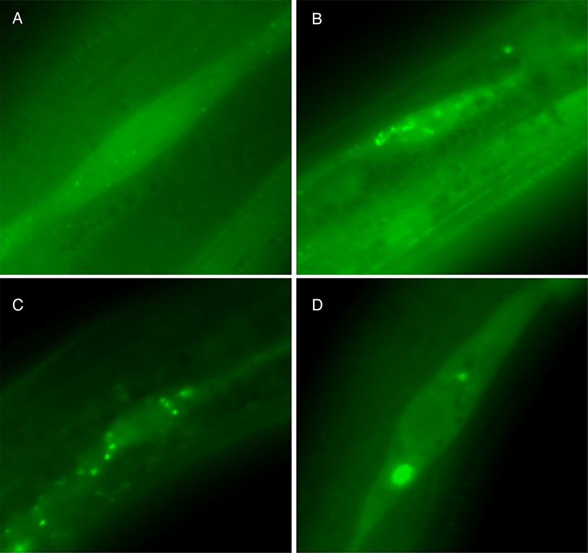
In yeast and mammalian cells, the autophagy protein Atg8/LC3 (microtubule-associated proteins 1A/1B light chain 3B encoded by MAP1LC3B) has been the marker of choice to detect double-membraned autophagosomes that are produced during the process of autophagy. A lipid-conjugated form of Atg8/LC3B is localized to the inner and outer membrane of the early-forming structure known as the phagophore. During maturation of autophagosomes, Atg8/LC3 bound to the inner autophagosome membrane remains in situ as the autophagosomes fuse with lysosomes. The nematode Caenorhabditis elegans is thought to conduct a similar process, meaning that tagging the nematode ortholog of Atg8/LC3-known as LGG-1-with a fluorophore has become a widely accepted method to visualize autophagosomes. Under normal growth conditions, GFP-modified LGG-1 displays a diffuse expression pattern throughout a variety of tissues, whereas, when under conditions that induce autophagy, the GFP::LGG-1 tag labels positive punctate structures, and its overall level of expression increases. Here, we present a protocol for using fluorescent reporters of LGG-1 coupled to GFP to monitor autophagosomes in vivo. We also discuss the use of alternative fluorescent markers and the possible utility of the LGG-1 paralog LGG-2.
© 2016 Cold Spring Harbor Laboratory Press.
Figures

Similar articles
-
Human GABARAP can restore autophagosome biogenesis in a C. elegans lgg-1 mutant.Autophagy. 2014 Oct 1;10(10):1868-72. doi: 10.4161/auto.29745. Epub 2014 Jul 23. Autophagy. 2014. PMID: 25126728 Free PMC article.
-
Detection of Autophagy in Caenorhabditis elegans by Western Blotting Analysis of LGG-1.Cold Spring Harb Protoc. 2016 Feb 1;2016(2):pdb.prot086512. doi: 10.1101/pdb.prot086512. Cold Spring Harb Protoc. 2016. PMID: 26832685 Free PMC article.
-
The C. elegans LC3 acts downstream of GABARAP to degrade autophagosomes by interacting with the HOPS subunit VPS39.Dev Cell. 2014 Jan 13;28(1):43-55. doi: 10.1016/j.devcel.2013.11.022. Epub 2013 Dec 26. Dev Cell. 2014. PMID: 24374177
-
Guidelines for monitoring autophagy in Caenorhabditis elegans.Autophagy. 2015;11(1):9-27. doi: 10.1080/15548627.2014.1003478. Autophagy. 2015. PMID: 25569839 Free PMC article. Review.
-
Autophagosomal Sperm Organelle Clearance and mtDNA Inheritance in C. elegans.Adv Anat Embryol Cell Biol. 2019;231:1-23. doi: 10.1007/102_2018_1. Adv Anat Embryol Cell Biol. 2019. PMID: 30467692 Review.
Cited by
-
Coupling of autophagy and the mitochondrial intrinsic apoptosis pathway modulates proteostasis and ageing in Caenorhabditis elegans.Cell Death Dis. 2023 Feb 11;14(2):110. doi: 10.1038/s41419-023-05638-x. Cell Death Dis. 2023. PMID: 36774344 Free PMC article.
-
Tripchlorolide induces autophagy in lung cancer cells by inhibiting the PI3K/AKT/mTOR pathway and improves cisplatin sensitivity in A549/DDP cells.Oncotarget. 2017 Jul 12;8(38):63911-63922. doi: 10.18632/oncotarget.19201. eCollection 2017 Sep 8. Oncotarget. 2017. PMID: 28969040 Free PMC article.
-
Acyl-CoA-binding protein (ACBP): a phylogenetically conserved appetite stimulator.Cell Death Dis. 2020 Jan 6;11(1):7. doi: 10.1038/s41419-019-2205-x. Cell Death Dis. 2020. PMID: 31907349 Free PMC article.
-
Neurotrophic factor MANF regulates autophagy and lysosome function to promote proteostasis in C. elegans.bioRxiv [Preprint]. 2024 Jan 4:2023.07.31.551399. doi: 10.1101/2023.07.31.551399. bioRxiv. 2024. Update in: Proc Natl Acad Sci U S A. 2024 Oct 22;121(43):e2403906121. doi: 10.1073/pnas.2403906121. PMID: 38260421 Free PMC article. Updated. Preprint.
-
Detection of Autophagy in Caenorhabditis elegans.Cold Spring Harb Protoc. 2016 Feb 1;2016(2):pdb.top070466. doi: 10.1101/pdb.top070466. Cold Spring Harb Protoc. 2016. PMID: 26832690 Free PMC article.
References
-
- Al Rawi S, Louvet-Vallee S, Djeddi A, Sachse M, Culetto E, Hajjar C, Boyd L, Legouis R, Galy V, 2011. Postfertilization autophagy of sperm organelles prevents paternal mitochondrial DNA transmission. Science 334, 1144–1147. - PubMed
-
- Albert PS, Brown SJ, Riddle DL, 1981. Sensory control of dauer larva formation in Caenorhabditis elegans. J Comp Neurol 198, 435–451. - PubMed
-
- Alberti A, Michelet X, Djeddi A, Legouis R, 2010. The autophagosomal protein LGG-2 acts synergistically with LGG-1 in dauer formation and longevity in C. elegans. Autophagy 6, 622–633. - PubMed
MeSH terms
Substances
Grants and funding
LinkOut - more resources
Full Text Sources
Other Literature Sources