. 2015 Oct;18(10):942-9.

Structural characterization of rat ventricular tissue exposed to the smoke of two types of waterpipe

Affiliations

Structural characterization of rat ventricular tissue exposed to the smoke of two types of waterpipe

Wajdy Al-Awaida et al. Iran J Basic Med Sci. 2015 Oct.

Abstract

Objectives: this study focused on the effect of waterpipe smoke exposure toxicity on the structure of albino rat's ventricular tissue and their recovery.

Materials and methods: Albino rats were divided into three groups: control, flavored, and unflavored. The control group was exposed to normal air while the flavored and unflavored groups were exposed to waterpipe smoke for a period of 90 days. Each group was followed by a period of 90 days of fresh air exposure. Following each period, the ventricular tissue was removed for biochemical and histopathological studies.

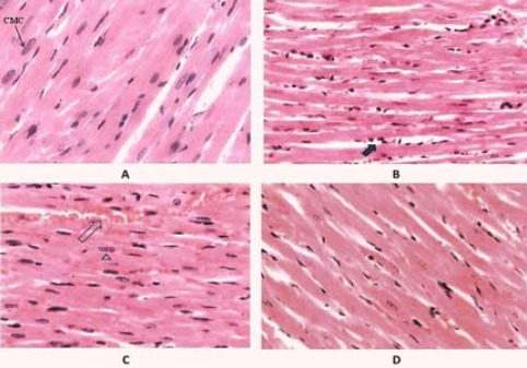
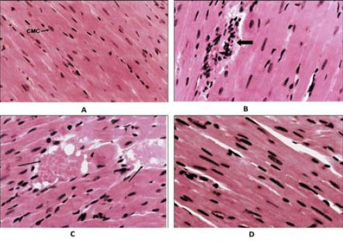
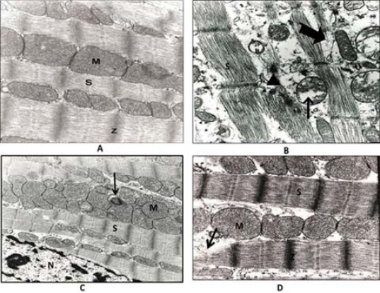
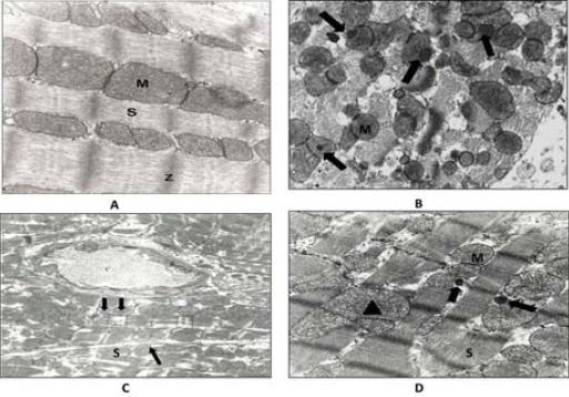
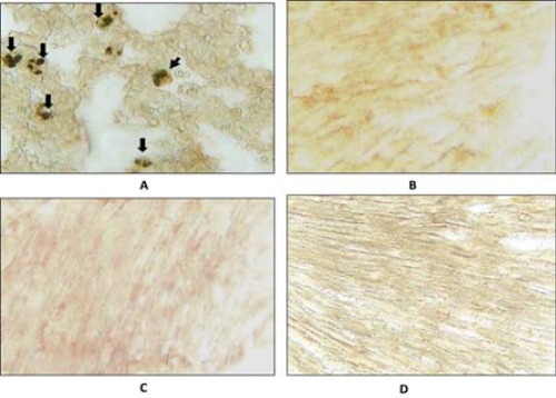
Results: The ventricular tissues of waterpipe exposed rats showed some degree of separation between cardiac muscle fibers, infiltration of lymphocytes, and congestion of blood vessel. Also, thin cross sections of ventricular cells revealed pleomorphic mitochondria with partially disrupted cristae, partial disruption of the myofibrils, and deposited toxic materials. The unflavored waterpipe has more deleterious effects on heart ventricular tissues than the flavored one. Waterpipe smoke didn't induce apoptosis in the ventricular tissue. We also found very high levels of plasma thiocyanate after exposure to smoke in the flavored and unflavored groups, while the control group showed no increase. After the recovery period, those tissues showed partial recovery.

Conclusion: Waterpipe smoke induces structural changes in the heart ventricle tissues, causing a negative impact on the capacity of the cardiac muscle for pumping blood and may lead to heart attack due to accumulation of free radicals and tissue inflammation. Cessation of smoking is important in returning most of these changes to their normal structure.

Keywords: Apoptosis; Thiocyanate level; Ultrastractural changes; Ventricular cell; Ventricular tissue; Waterpipe smoke.


Figures

Figure 1
Figure 1
Effect of flavored waterpipe exposed rat on heart ventricular tissue using hematoxylin and eosin staining. A: normal morphology of heart ventricular tissue of air-exposed rat. CMC: cardiac muscle cell. Magnification: 830x. B: heart ventricular tissue of flavored waterpipe exposed rat, showing some degree of separation between cardiac muscle fibers. The arrow indicates lymphocytic infiltration. Magnification: 540x. C: heart ventricular tissue of flavored waterpipe exposed rat. Arrow indicates congested blood vessel. Magnification: 630x. D: Heart ventricular tissue of flavored waterpipe exposed rat after the recovery period. The tissue almost returned to its normal appearance. Magnification: 540x
Figure 2
Figure 2
The effect of unflavored waterpipe exposed rat on heart ventricular tissue using hematoxylin and eosin staining. A: normal morphology of heart ventricular tissue of air-exposed rat. CMC: cardiac muscle cell. Magnification: 830x. B: heart ventricular tissue of unflavored waterpipe exposed rat. Arrow indicates lymphocytic infiltration. Magnification: 790x. C: heart ventricular tissue of unflavored waterpipe exposed rat. Arrow indicates congested blood vessel. Magnification: 760x. D: heart ventricular tissue of flavored waterpipe exposed rat after the recovery period. The tissue almost returned to its normal appearance. Magnification: 1130x
Figure 3
Figure 3
Effect of flavored waterpipe exposed rat on heart ventricular tissue. A: ultrathin section of ventricular cardiomyocytes of air-exposed rat. M: mitochondrion with normal cristae, S: sarcomeres, Z: Z-line. Magnification:62500x. B: ultrathin section of ventricular cardiomyocytes of flavored waterpipe exposed rat. The thick arrows indicate a partial disruption of the myofibrils. Thin arrow indicates pleomorphic mitochondria with partially disrupted cristae. The triangle indicates deposited material. Magnification: 62500x. C: Ultrathin section of ventricular cardiomyocytes of flavored waterpipe exposed rat, showing enlarged mitochondria, irregularly arranged with partial disrupted cristae. Arrow indicates deposited material, M: mitochondrion, S: sarcomeres, N: nucleus. Magnification:25000x. D: ultrathin section of ventricular cardiomyocytes of flavored waterpipe exposed rat after the recovery period, showing partial recovery of cardiac muscle fiber. Enlarged mitochondria with partial disruption cristae. Arrow indicates a partial disruption of the myofibrils, M: mitochondrion, S: sarcomere. Magnification:62500x
Figure 4
Figure 4
Effect of unflavored waterpipe exposed rat on heart ventricular tissue. A: ultrathin section of ventricular cardiomyocytes of air-exposed rat. M: mitochondrion with normal cristae, S: sarcomeres, Z: Z-line. Magnification:62500x. B: ultrathin section of ventricular cells of unflavored waterpipe exposed rat, showing pleomorphic mitochondria. Arrows indicate deposited material. Magnification 5000x. C: ultrathin section of ventricular cells of flavored waterpipe exposed rat. The two areas enclosed by trapezoidal shapes denote partial disruption of the Z-disc. Arrows indicate partially disrupted mitochondria. C: capillary. Magnification: 5000x. D: ultrathin section of ventricular cells of unflavored waterpipe exposed rat after the recovery period, showing partial recovery of cardiac muscle fiber. Arrows indicate deposited material. The triangle indicates enlarged and pleomorphic mitochondria with partially disrupted cristae. M: mitochondrion, S: sarcomere Magnification: 25000x
Figure 5
Figure 5
The ability of waterpipe smoke (flavored and unflavored) to induce apoptosis in heart ventricular tissue. A: positive control, with the presence of many dark-brown apoptotic nuclei. Arrows indicate one apoptotic nucleus (TUNEL). B: heart ventricular tissue of air-exposed rat, no dark-brown apoptotic nuclei. C: heart ventricular tissue of flavored waterpipe exposed rat, no dark-brown apoptotic nuclei. D: heart ventricular tissue of unflavored waterpipe exposed rat, no dark-brown apoptotic nuclei

References

    1. Hoffmann D, Wynder EL. Chemical constituents and bioactivity of tobacco smoke. IARC Sci Publ. 1986;74:145–165. - PubMed
    1. Chaouachi K. Hookah (Shisha, Narghile) smoking and environmental tobacco smoke (ETS), a critical review of the relevant literature and the public health consequences. Int J Environ Res Publ Health. 2009;6:798–843. - PMC - PubMed
    1. Chaouachi K. Public health intervention for narghile (hookah, shisha) use requires a radical critique of the related “standardised” smoking machine. J Public Health. 2010;18:69–73.
    1. Monzer B, Sepetdjian E, Saliba N, Shihadeh A. Charcoal emissions as a source of CO and carcinogenic PAH in mainstream narghile waterpipe smoke. Food Chem Toxicol. 2008;46:2991–2995. - PubMed
    1. Shihadeh A. Investigation of the mainstream smoke aerosol of the argileh water pipe. Food Chem Toxicol. 2003;41:143–152. - PubMed

LinkOut - more resources