

Cephalometric landmark variability among orthodontists and dentomaxillofacial radiologists: a comparative study
- PMID: 26730368
- PMCID: PMC4697005
- DOI: 10.5624/isd.2015.45.4.213
Cephalometric landmark variability among orthodontists and dentomaxillofacial radiologists: a comparative study
Abstract
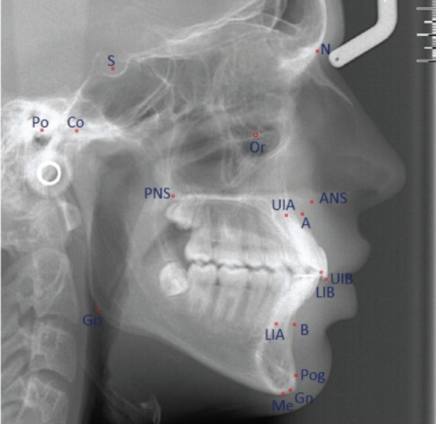
Purpose: The aim this study was to compare the accuracy of orthodontists and dentomaxillofacial radiologists in identifying 17 commonly used cephalometric landmarks, and to determine the extent of variability associated with each of those landmarks.
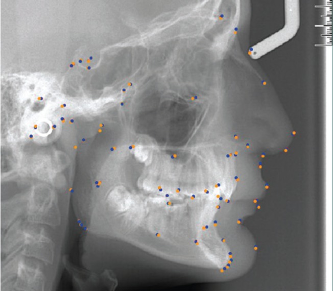
Materials and methods: Twenty digital lateral cephalometric radiographs were evaluated by two groups of dental specialists, and 17 cephalometric landmarks were identified. The x and y coordinates of each landmark were recorded. The mean value for each landmark was considered the best estimate and used as the standard. Variation in measurements of the distance between landmarks and measurements of the angles associated with certain landmarks was also assessed by a subset of two observers, and intraobserver and interobserver agreement were evaluated.
Results: Intraclass correlation coefficients were excellent for intraobserver agreement, but only good for interobserver agreement. The least reliable landmark for orthodontists was the gnathion (Gn) point (standard deviation [SD], 5.92 mm), while the orbitale (Or) was the least reliable landmark (SD, 4.41 mm) for dentomaxillofacial radiologists. Furthermore, the condylion (Co)-Gn plane was the least consistent (SD, 4.43 mm).
Conclusion: We established that some landmarks were not as reproducible as others, both horizontally and vertically. The most consistently identified landmark in both groups was the lower incisor border, while the least reliable points were Co, Gn, Or, and the anterior nasal spine. Overall, a lower level of reproducibility in the identification of cephalometric landmarks was observed among orthodontists.
Keywords: Analysis; Anatomic Landmarks; Cephalometry; Orthodontics; Reliability and Validity.
Figures
References
-
- Broadbent BH. A new x-ray technique and its application to orthodontia. Angle Orthod. 1931;1:45–66.
-
- Baumrind S, Frantz RC. The reliability of head film measurements. 1. Landmark identification. Am J Orthod. 1971;60:111–127. - PubMed
-
- Chen YJ, Chen SK, Huang HW, Yao CC, Chang HF. Reliability of landmark identification in cephalometric radiography acquired by a storage phosphor imaging system. Dentomaxillofac Radiol. 2004;33:301–306. - PubMed
-
- Kamoen A, Dermaut L, Verbeeck R. The clinical significance of error measurement in the interpretation of treatment results. Eur J Orthod. 2001;23:569–578. - PubMed
-
- Miloro M, Borba AM, Ribeiro-Junior O, Naclério-Homem MG, Jungner M. Is there consistency in cephalometric landmark identification amongst oral and maxillofacial surgeons? Int J Oral Maxillofac Surg. 2014;43:445–453. - PubMed
LinkOut - more resources
Full Text Sources
Other Literature Sources
