

Assessment of maxillary third molars with panoramic radiography and cone-beam computed tomography
- PMID: 26730371
- PMCID: PMC4697008
- DOI: 10.5624/isd.2015.45.4.233
Assessment of maxillary third molars with panoramic radiography and cone-beam computed tomography
Abstract
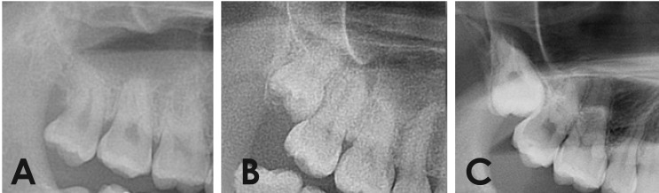
Purpose: This study investigated maxillary third molars and their relation to the maxillary sinus using panoramic radiography and cone-beam computed tomography (CBCT).
Materials and methods: A total of 395 maxillary third molars in 234 patients were examined using panoramic radiographs and CBCT images. We examined the eruption level of the maxillary third molars, the available retromolar space, the angulation, the relationship to the second molars, the number of roots, and the relationship between the roots and the sinus.
Results: Females had a higher frequency of maxillary third molars with occlusal planes apical to the cervical line of the second molar (Level C) than males. All third molars with insufficient retromolar space were Level C. The most common angulation was vertical, followed by buccoangular. Almost all of the Level C molars were in contact with the roots of the second molar. Erupted teeth most commonly had three roots, and completely impacted teeth most commonly had one root. The superimposition of one third of the root and the sinus floor was most commonly associated with the sinus floor being located on the buccal side of the root.
Conclusion: Eruption levels were differently distributed according to gender. A statistically significant association was found between the eruption level and the available retromolar space. When panoramic radiographs showed a superimposition of the roots and the sinus floor, expansion of the sinus to the buccal side of the root was generally observed in CBCT images.
Keywords: Cone-Beam Computed Tomography; Maxilla; Maxillary sinus; Molar, Third; Radiography, Panoramic.
Figures


References
-
- de Carvalho RW, de Araújo Filho RC, do Egito Vasconcelos BC. Assessment of factors associated with surgical difficulty during removal of impacted maxillary third molars. J Oral Maxillofac Surg. 2013;71:839–845. - PubMed
-
- Patel M, Down K. Accidental displacement of impacted maxillary third molars. Br Dent J. 1994;177:57–59. - PubMed
-
- Dimitrakopoulos I, Papadaki M. Displacement of a maxillary third molar into the infratemporal fossa: case report. Quintessence Int. 2007;38:607–610. - PubMed
-
- Gómez-Oliveira G, Arribas-García I, Alvarez-Flores M, Gregoire-Ferriol J, Martínez-Gimeno C. Delayed removal of a maxillary third molar from the infratemporal fossa. Med Oral Patol Oral Cir Bucal. 2010;15:e509–e511. - PubMed
-
- Oberman M, Horowitz I, Ramon Y. Accidental displacement of impacted maxillary third molars. Int J Oral Maxillofac Surg. 1986;15:756–758. - PubMed
LinkOut - more resources
Full Text Sources
Other Literature Sources
