. 2015 Oct;2(4):041407.
doi: 10.1117/1.NPh.2.4.041407. Epub 2015 Nov 9.

Ultramicroscopy: development and outlook

Affiliations

Ultramicroscopy: development and outlook

Hans-Ulrich Dodt et al. Neurophotonics. 2015 Oct.

Abstract

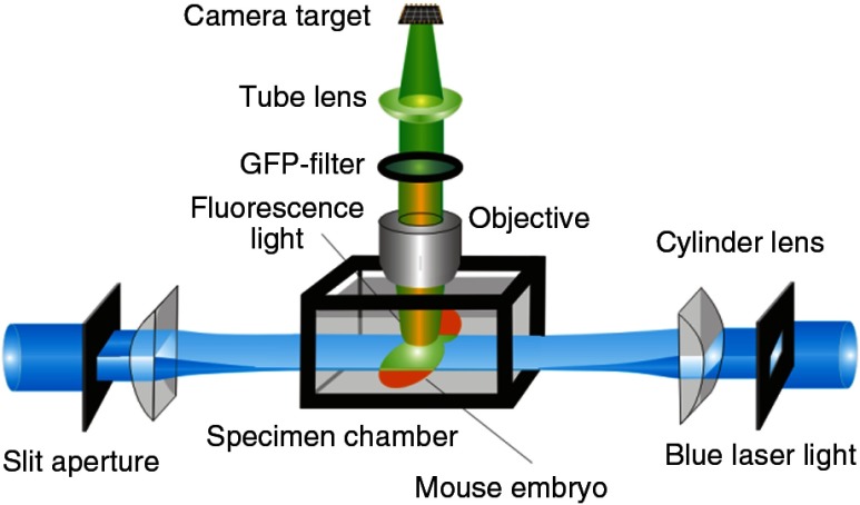
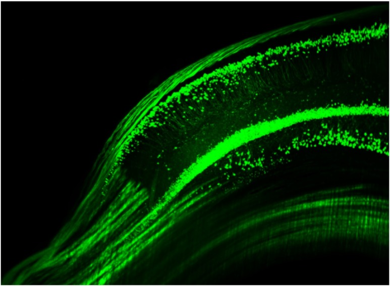
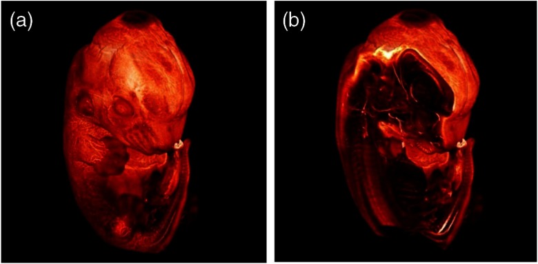
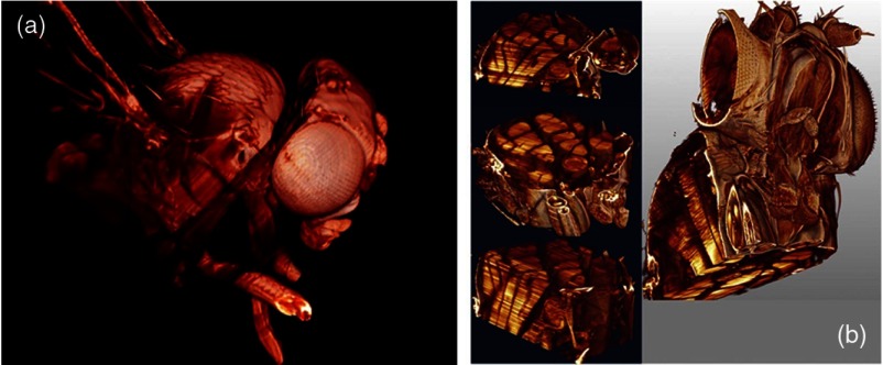
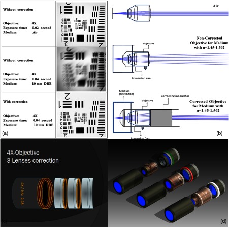
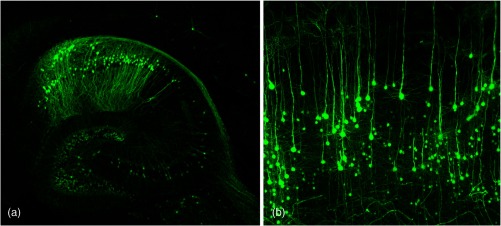
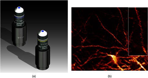
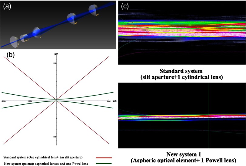
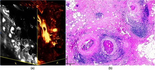
We present an overview of the ultramicroscopy technique we developed. Starting from developments 100 years ago, we designed a light sheet microscope and a chemical clearing to image complete mouse brains. Fluorescence of green fluorescent protein (GFP)-labeled neurons in mouse brains could be preserved with our 3DISCO clearing and high-resolution three-dimensional (3-D) recordings were obtained. Ultramicroscopy was also used to image whole mouse embryos and flies. We improved the optical sectioning of our light sheet microscope by generating longer and thinner light sheets with aspheric optics. To obtain high-resolution images, we corrected available air microscope objectives for clearing solutions with high refractive index. We discuss how eventually super resolution could be realized in light sheet microscopy by applying stimulated emission depletion technology. Also the imaging of brain function by recording of mouse brains expressing cfos-GFP is discussed. Finally, we show the first 3-D recordings of human breast cancer with light sheet microscopy as application in medical diagnostics.

Keywords: brain clearing; light sheet; objective correction; three-dimensional brain imaging; tumor clearing; ultramicroscopy.


Figures

Fig. 1
Fig. 1
Schematic drawing of the optics of an ultramicroscope.
Fig. 2
Fig. 2
Green fluorescent protein (GFP)-labeled neurons in the hippocampus.
Fig. 3
Fig. 3
Three-dimensional (3-D) reconstruction of a mouse embryo. (a) The surface rendering shows the external view and (b) nervous structures inside the embryo made visible with fluorescent antibody staining.
Fig. 4
Fig. 4
3-D reconstruction of Drosophila melanogaster: (a) surface and (b) internal features clearly depicting flight muscles.
Fig. 5
Fig. 5
(a) Images obtained with 4× objective (NA, 0.28) in air and DBE without and with correction, (b) optical arrangement for objective (without and with correction optics), (c) 4× objective with correction optics, and (d) three corrected air objectives, 2× (NA, 0.14), 4× (NA, 0.28) and 20× (NA, 0.45).
Fig. 6
Fig. 6
(a) Anatomy of the hippocampus (20 optical sections) imaged in the whole mouse brain with a corrected 4× objective (NA, 0.28). (b) Neurons in the neocortex imaged with corrected 4× objective (300 optical sections).
Fig. 7
Fig. 7
(a) 40× (NA, 0.8) and 10× (NA, 0.3) water immersion objectives corrected for n=1.45 (CLARITY). (b) Already with the corrected 10× objective, spines become visible in a CLARITY-cleared whole mouse brain.
Fig. 8
Fig. 8
(a) Light sheet-generating optics, (b) profile of light sheets obtained with two different optical systems: red Gaussian, green generated with one Powell lens, and (c) comparison of the focus of the two light sheets.
Fig. 9
Fig. 9
Human breast cancer (DCIS) imaged with a 2× objective. Both in the 3-D reconstruction (a) and the standard histological section and (b) the clogged milk-duct-like structures can be seen.

References

    1. Helmchen F., Denk W., “Deep tissue two-photon microscopy,” Nat. Methods 2, 932–940 (2005).10.1038/nmeth818 - DOI - PubMed
    1. Johnson G. A., et al. , “Morphological phenotyping with MR microscopy: the visible mouse,” Radiology 222, 789–793 (2002).RADLAX10.1148/radiol.2223010531 - DOI - PubMed
    1. Kalender W. A., “CT: the unexpected evolution of an imaging modality,” Eur Radiol. Suppl. 15(Suppl. 4), D21–D24 (2005).10.1007/s10406-005-0128-3 - DOI - PubMed
    1. Tyszka J. M., Fraser S. E., Jacobs R. E., “Magnetic resonance microscopy: recent advances and applications,” Curr. Opin. Biotechnol. 16, 93–99 (2005).CUOBE310.1016/j.copbio.2004.11.004 - DOI - PubMed
    1. Ragan T., et al. , “High-resolution whole organ imaging using two-photon tissue cytometry,” J. Biomed. Opt. 12, 014015 (2007).10.1117/1.2435626 - DOI - PubMed