

Calcium Signaling Is Required for Erythroid Enucleation
- PMID: 26731108
- PMCID: PMC4701494
- DOI: 10.1371/journal.pone.0146201
Calcium Signaling Is Required for Erythroid Enucleation
Abstract
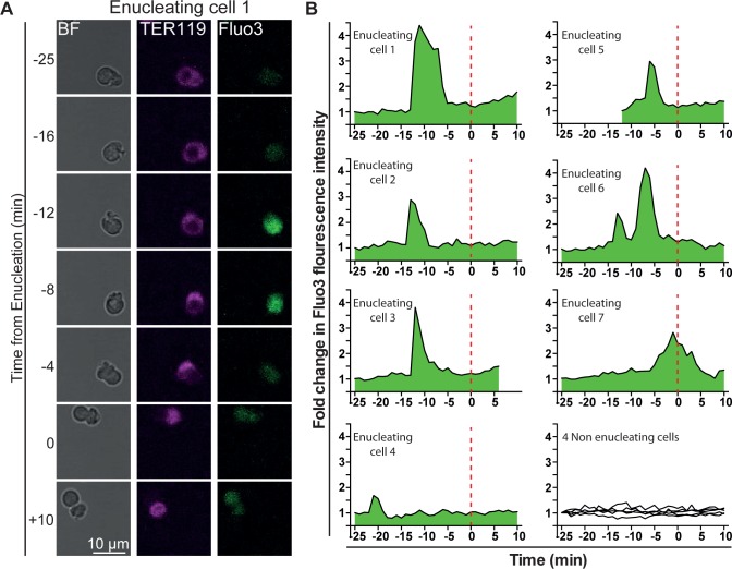
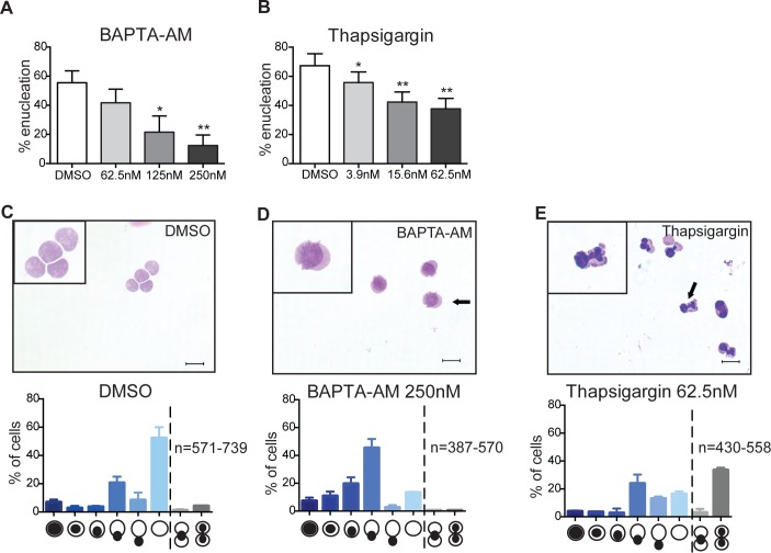
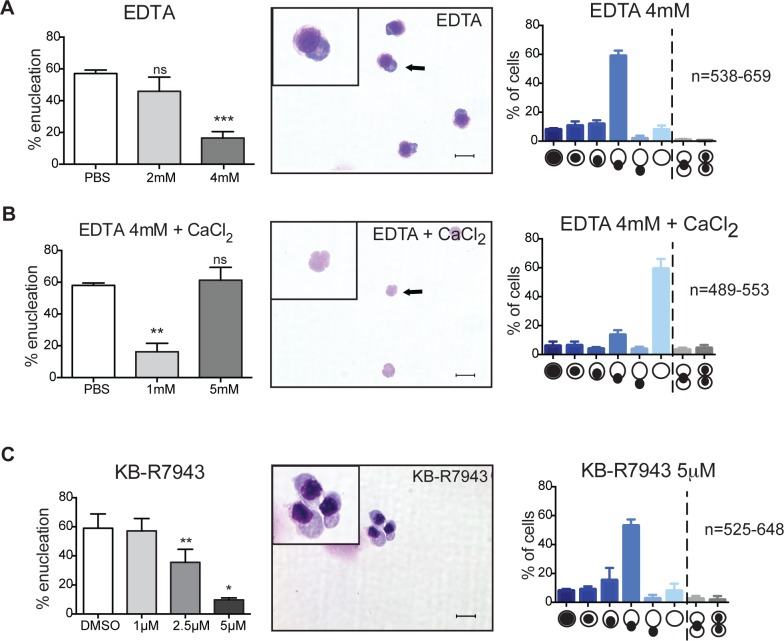
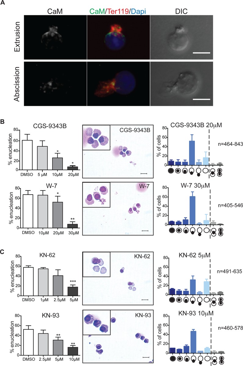
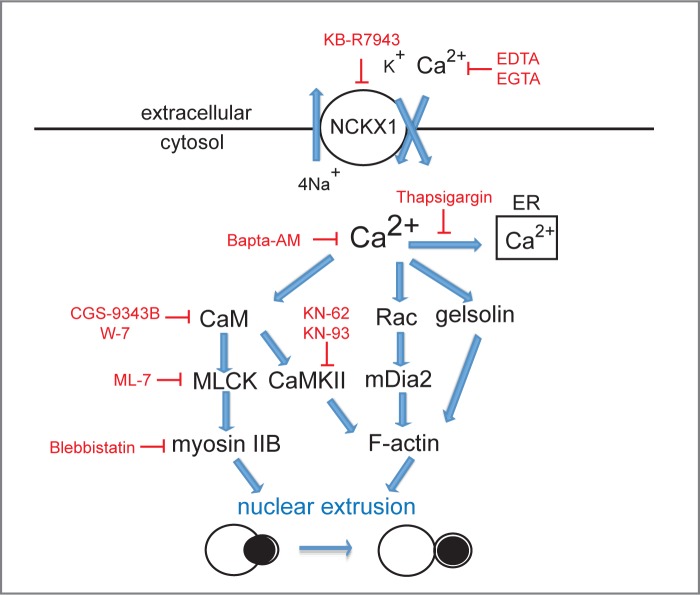
Although erythroid enucleation, the property of erythroblasts to expel their nucleus, has been known for 7ore than a century, surprisingly little is known regarding the molecular mechanisms governing this unique developmental process. Here we show that similar to cytokinesis, nuclear extrusion requires intracellular calcium signaling and signal transduction through the calmodulin (CaM) pathway. However, in contrast to cytokinesis we found that orthochromatic erythroblasts require uptake of extracellular calcium to enucleate. Together these functional studies highlight a critical role for calcium signaling in the regulation of erythroid enucleation.
Conflict of interest statement
Figures
Similar articles
-
A Chemical Screening Approach to Identify Novel Key Mediators of Erythroid Enucleation.PLoS One. 2015 Nov 16;10(11):e0142655. doi: 10.1371/journal.pone.0142655. eCollection 2015. PLoS One. 2015. PMID: 26569102 Free PMC article.
-
Fine-tuned calcium homeostasis is crucial for murine erythropoiesis.FEBS J. 2025 Apr;292(8):1934-1949. doi: 10.1111/febs.17401. Epub 2025 Jan 21. FEBS J. 2025. PMID: 39838539
-
The Asymmetric Cell Division Regulators Par3, Scribble and Pins/Gpsm2 Are Not Essential for Erythroid Development or Enucleation.PLoS One. 2017 Jan 17;12(1):e0170295. doi: 10.1371/journal.pone.0170295. eCollection 2017. PLoS One. 2017. PMID: 28095473 Free PMC article.
-
Rho GTPases in erythroid maturation.Curr Opin Hematol. 2014 May;21(3):165-71. doi: 10.1097/MOH.0000000000000032. Curr Opin Hematol. 2014. PMID: 24492678 Free PMC article. Review.
-
Understanding terminal erythropoiesis: An update on chromatin condensation, enucleation, and reticulocyte maturation.Blood Rev. 2021 Mar;46:100740. doi: 10.1016/j.blre.2020.100740. Epub 2020 Aug 8. Blood Rev. 2021. PMID: 32798012 Review.
Cited by
-
Erythroblast enucleation at a glance.J Cell Sci. 2024 Oct 1;137(19):jcs261673. doi: 10.1242/jcs.261673. Epub 2024 Oct 14. J Cell Sci. 2024. PMID: 39397781 Free PMC article. Review.
-
Biogenesis and Breakdown of Lipid Droplets in Pathological Conditions.Front Cell Dev Biol. 2022 Feb 7;9:826248. doi: 10.3389/fcell.2021.826248. eCollection 2021. Front Cell Dev Biol. 2022. PMID: 35198567 Free PMC article. Review.
-
Dysregulation of erythropoiesis and altered erythroblastic NMDA receptor-mediated calcium influx in Lrfn2-deficient mice.PLoS One. 2021 Jan 22;16(1):e0245624. doi: 10.1371/journal.pone.0245624. eCollection 2021. PLoS One. 2021. PMID: 33481887 Free PMC article.
-
Cellular dynamics of mammalian red blood cell production in the erythroblastic island niche.Biophys Rev. 2019 Dec;11(6):873-894. doi: 10.1007/s12551-019-00579-2. Epub 2019 Aug 15. Biophys Rev. 2019. PMID: 31418139 Free PMC article. Review.
-
PIEZO1 activation delays erythroid differentiation of normal and hereditary xerocytosis-derived human progenitor cells.Haematologica. 2020 Mar;105(3):610-622. doi: 10.3324/haematol.2019.218503. Epub 2019 Aug 14. Haematologica. 2020. PMID: 31413092 Free PMC article.
References
Publication types
MeSH terms
Substances
LinkOut - more resources
Full Text Sources
Other Literature Sources
