. 2016 Feb;126(2):706-20.
doi: 10.1172/JCI78132. Epub 2016 Jan 5.

Histone demethylase JMJD2A drives prostate tumorigenesis through transcription factor ETV1

Histone demethylase JMJD2A drives prostate tumorigenesis through transcription factor ETV1

Tae-Dong Kim et al. J Clin Invest. 2016 Feb.

Abstract

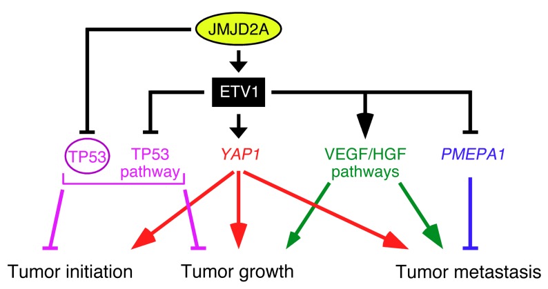
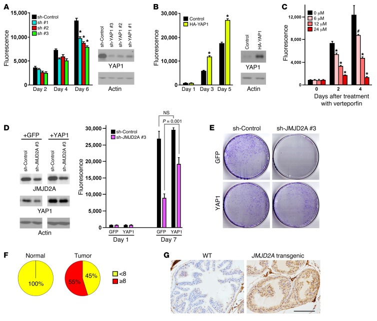
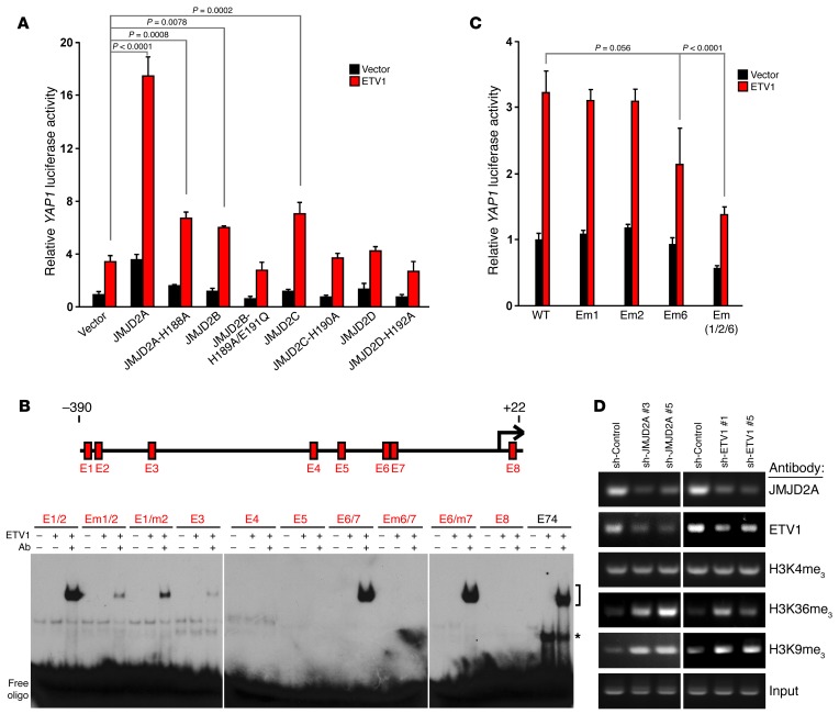
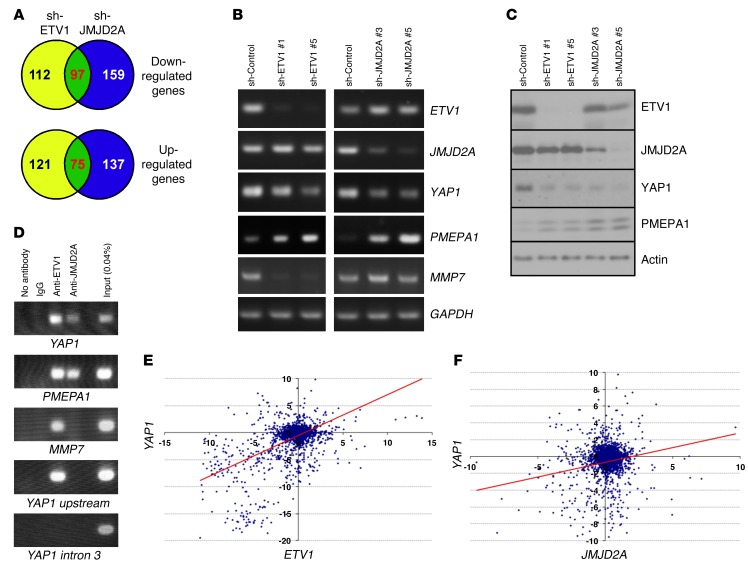
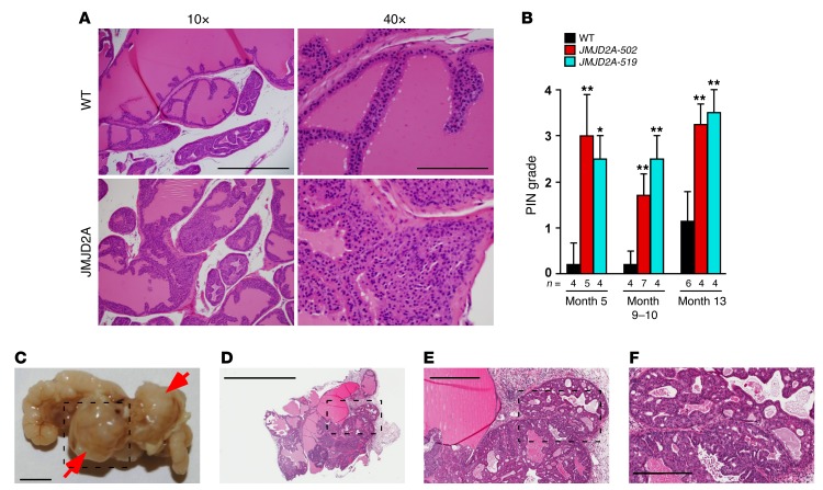
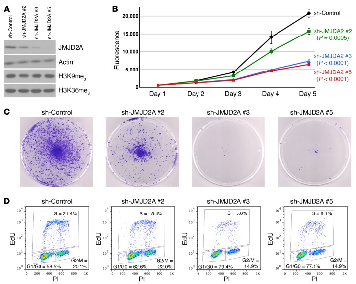
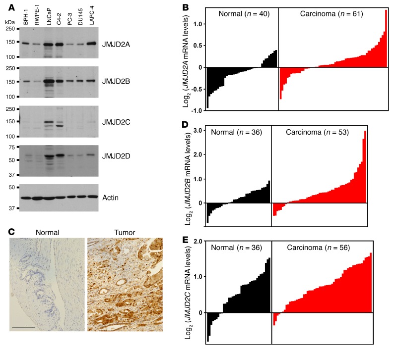
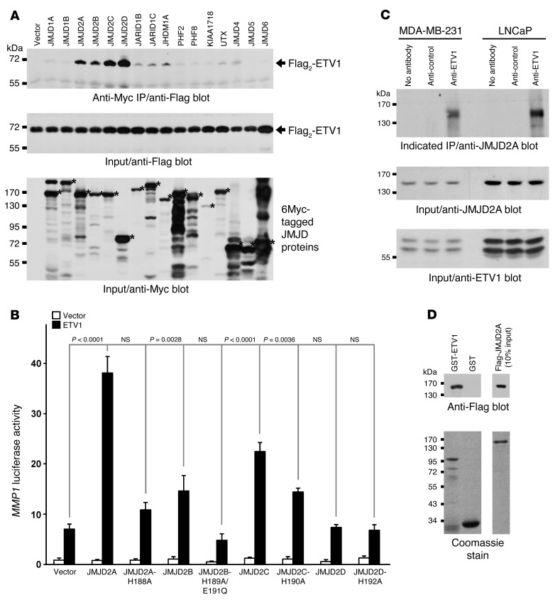
Histone demethylase upregulation has been observed in human cancers, yet it is unknown whether this is a bystander event or a driver of tumorigenesis. We found that overexpression of lysine-specific demethylase 4A (KDM4A, also known as JMJD2A) was positively correlated with Gleason score and metastasis in human prostate tumors. Overexpression of JMJD2A resulted in the development of prostatic intraepithelial neoplasia in mice, demonstrating that JMJD2A can initiate prostate cancer development. Moreover, combined overexpression of JMJD2A and the ETS transcription factor ETV1, a JMJD2A-binding protein, resulted in prostate carcinoma formation in mice haplodeficient for the phosphatase and tensin homolog (Pten) tumor-suppressor gene. Additionally, JMJD2A cooperated with ETV1 to increase expression of yes associated protein 1 (YAP1), a Hippo pathway component that itself was associated with prostate tumor aggressiveness. ETV1 facilitated the recruitment of JMJD2A to the YAP1 promoter, leading to changes in histone lysine methylation in a human prostate cancer cell line. Further, YAP1 expression largely rescued the growth inhibitory effects of JMJD2A depletion in prostate cancer cells, indicating that YAP1 is a downstream effector of JMJD2A. Taken together, these data reveal a JMJD2A/ETV1/YAP1 axis that promotes prostate cancer initiation and that may be a suitable target for therapeutic inhibition.


Figures

Figure 8
Figure 8. Model.
Pleiotropic mechanisms by which JMJD2A promotes tumorigenesis.
Figure 7
Figure 7. Role of YAP1 in prostate cancer.
(A) Downregulation of YAP1 reduces and (B) overexpression of YAP1 stimulates LNCaP cell growth. Representative Western blots of shRNA-mediated knockdown of YAP1 (A) and HA-YAP1 expression (B) are shown. *P < 0.0001, 1-way ANOVA (A); Student’s t test (B). Shown are averages (n = 3 per group) with SD. Representative of 3 independent experiments. (C) LNCaP cell growth after addition of 0–24 μM verteporfin dissolved in DMSO (20 mM stock). #P = 0.0002; *P < 0.0001 (1-way ANOVA). Shown are averages (n = 4 per group) with SD. Representative of 2 independent experiments. (D) Ectopic YAP1, but not GFP, counteracts LNCaP growth inhibition caused by JMJD2A shRNA. Shown are averages (n = 3 per group) with SD. 1-way ANOVA. Representative of 2 independent experiments. (E) Likewise, YAP1 rescues LNCaP clonogenic capacity upon JMJD2A downregulation (representative out of 3 experiments). (F) Nuclear YAP1 overexpression in 31 cancerous prostate versus matching normal tissues (staining index < 8 and ≥ 8; P = 7.2 × 10⁻⁷, 2-tailed Fisher exact probability test). (G) Example of YAP1 immunohistochemical staining in prostates from a WT or JMJD2A transgenic mouse. Scale bar: 0.1 mm.
Figure 6
Figure 6. Characterization of the YAP1 promoter.
(A) Activation of a YAP1 promoter (–390/+22) luciferase reporter by ETV1 and JMJD2 protein (or respective catalytic mutant) in LNCaP cells. Shown are averages (n = 3 biological replicates per group) with SD and 1-way ANOVA probabilities. (B) Electrophoretic mobility shift assays with 32P-labeled oligonucleotides corresponding to the 8 ETS sites (E1 to E8) within the YAP1 promoter or the E74 binding site. m, mutated ETS site; asterisk denotes an ETV1:DNA complex, whereas the bracket marks the supershift of ETV1:DNA complexes caused by an ETV1 Ab. (C) Impact of mutating E1, E2, and/or E6 on YAP1 promoter luciferase activity in LNCaP cells. Shown are averages (n = 3 biological replicates per group) with SD. Significance was determined with 1-way ANOVA. (D) YAP1 promoter ChIP assays with indicated Abs on LNCaP cells treated with JMJD2A or ETV1 shRNA. Amplified DNA fragments covered the promoter from –440 to –12.
Figure 5
Figure 5. Identification of JMJD2A/ETV1 target genes.
(A) Venn diagrams of genes down- or upregulated upon treatment of LNCaP cells with ETV1 or JMJD2A shRNAs. (B) Validation of target genes by RT-PCR in LNCaP cells. (C) Corresponding analysis of protein levels. (D) ChIP assay with ETV1 and JMJD2A Abs at indicated gene promoters in LNCaP cells. YAP1 upstream is approximately 2.5 kb upstream, and YAP1 intron 3 is approximately 72 kb downstream of the YAP1 transcription start site. (E) Correlation between fold changes in YAP1 and ETV1 mRNA levels across 3,949 microarrays. Pearson correlation: R = 0.52; P = 4.64 × 10–177. (F) Correlation between fold changes in YAP1 and JMJD2A mRNA levels across 3,949 microarrays. Pearson correlation: R = 0.12; P = 2.29 × 10⁻¹⁰.
Figure 4
Figure 4. Prostate phenotypes of transgenic JMJD2A mice.
(A) H&E staining of a sectioned prostate (anterior lobe) from a WT or JMJD2A transgenic mouse. Scale bars: 0.5 mm (left); 0.125 mm (right). (B) Summary of PIN formation in WT mice and 2 independent transgenic JMJD2A lines (502 and 519). Shown are PIN grade averages with SD and number of animals at 3 different ages. *P < 0.005; **P < 0.001, 1-way ANOVA. (C) Prostate with bladder and seminal vesicles from a JMJD2A/ETV1/Pten+/– mouse; arrows point to tumor masses. Scale bar: 5 mm. (DF) Progressively higher magnifications of H&E-stained sections of the lower tumor mass shown in C. Scale bars: 3 mm (D); 0.6 mm (E); 0.3 mm (F).
Figure 3
Figure 3. Role of JMJD2A in LNCaP prostate cancer cells.
(A) Downregulation of JMJD2A in LNCaP cells with 3 different shRNAs. Western blots show levels of indicated proteins. (B) Corresponding cell growth assays. Shown are averages (n = 3 per group) with SD. Statistical significance of growth differences (compared with sh-Control) at day 5 was evaluated with 1-way ANOVA. Representative of at least 3 independent experiments. (C) Representative (out of 3) clonogenic assays. (D) Cell-cycle distribution (representative out of 2 independent experiments) in the presence of JMJD2A shRNA.
Figure 2
Figure 2. JMJD2 expression in the human prostate.
(A) Western blots showing JMJD2 protein levels in untransformed (BPH-1, RWPE-1) and cancerous prostate cells (LNCaP, C4-2, PC-3, DU145, LAPC-4). (B) Different JMJD2A mRNA levels in 40 normal compared with 61 cancerous prostate tissues (P = 3.8 × 10⁻⁵; Student’s t test). Each bar represents 1 patient. (C) Example of immunohistochemical JMJD2A staining in matching normal and cancerous prostate tissue. Scale bar: 0.1 mm. (D and E) Analogous to panel B for JMJD2B (P = 0.003) and JMJD2C (P = 0.021).
Figure 1
Figure 1. Interaction of JMJD2A with ETV1.
(A) Coimmunoprecipitation assays of Flag2-ETV1 with 16 different 6Myc-tagged JMJD proteins (denoted by asterisks) in 293T cells. (B) Activation of an MMP1 luciferase reporter in BPH-1 cells. Luciferase activities shown are the average of 3 biological replicates with SD evaluated with 1-way ANOVA. (C) JMJD2A coprecipitates with ETV1 in MDA-MB-231 and LNCaP cells. (D) In vitro interaction of purified, Flag-tagged JMJD2A with GST-ETV1 (top). Coomassie-stained gels show the purity and relative amounts of utilized proteins (bottom).

References

    1. Ryan CJ, Tindall DJ. Androgen receptor rediscovered: the new biology and targeting the androgen receptor therapeutically. J Clin Oncol. 2011;29(27):3651–3658. doi: 10.1200/JCO.2011.35.2005. - DOI - PubMed
    1. Shen MM, Abate-Shen C. Molecular genetics of prostate cancer: new prospects for old challenges. Genes Dev. 2010;24(18):1967–2000. doi: 10.1101/gad.1965810. - DOI - PMC - PubMed
    1. Clark JP, Cooper CS. ETS gene fusions in prostate cancer. Nat Rev Urol. 2009;6(8):429–439. doi: 10.1038/nrurol.2009.127. - DOI - PubMed
    1. Hollenhorst PC, McIntosh LP, Graves BJ. Genomic and biochemical insights into the specificity of ETS transcription factors. Annu Rev Biochem. 2011;80:437–471. doi: 10.1146/annurev.biochem.79.081507.103945. - DOI - PMC - PubMed
    1. Oh S, Shin S, Janknecht R. ETV1, 4 and 5: An oncogenic subfamily of ETS transcription factors. Biochim Biophys Acta. 2012;1826(1):1–12. - PMC - PubMed

Publication types

MeSH terms