

In Vitro Analysis of Fibronectin-Modified Titanium Surfaces
- PMID: 26731536
- PMCID: PMC4711664
- DOI: 10.1371/journal.pone.0146219
In Vitro Analysis of Fibronectin-Modified Titanium Surfaces
Abstract
Background: Glow discharge plasma (GDP) procedure is an effective method for grafting various proteins, including albumin, type I collagen, and fibronectin, onto a titanium surface. However, the behavior and impact of titanium (Ti) surface modification is yet to be unraveled.
Purpose: The purpose of this study is to evaluate and analyze the biological properties of fibronectin-grafted Ti surfaces treated by GDP.
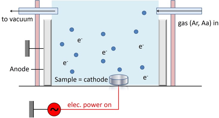
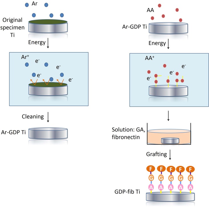
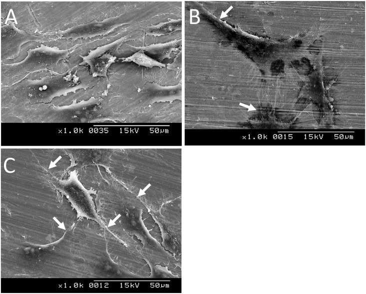
Materials and methods: Grade II Ti discs were initially cleaned and autoclaved to obtain original specimens. Subsequently, the specimens were GDP treated and grafted with fibronectin to form Ar-GDP (Argon GDP treatment only) and GDP-fib (fibronectin coating following GDP treatment) groups. Blood coagulation test and MG-63 cell culture were performed to evaluate the biological effects on the specimen.
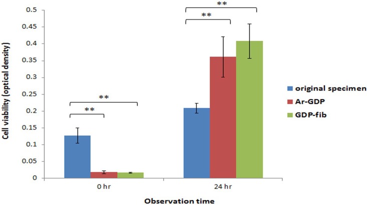
Results: There was no significant difference between Ar-GDP and GDP-fib groups in blood compatibility analysis. While in the MTT test, cellular proliferation was benefited from the presence of fibronectin coating. The numbers of cells on Ar-GDP and GDP-fib specimens were greater than those in the original specimens after 24 h of culturing.
Conclusions: GDP treatment combined with fibronectin grafting favored MG-63 cell adhesion, migration, and proliferation on titanium surfaces, which could be attributed to the improved surface properties.
Conflict of interest statement
Figures

References
-
- Branemark P.I. Osseointegration and its experimental background. J Prosthet Dent 1983, 50, 399–410. - PubMed
-
- Wennerberg A.; Hallgren C.; Johansson C.; Danelli S. A histomorphometric evaluation of screw-shaped implants each prepared with two surface roughnesses. Clin Oral Implants Res 1998, 9, 11–19. - PubMed
-
- Lazzara R.J.; Testori T.; Trisi P.; Porter S.S.; Weinstein R.L. A human histologic analysis of osseotite and machined surfaces using implants with 2 opposing surfaces. Int J Periodontics Restorative Dent 1999, 19, 117–129. - PubMed
-
- Oshida Y.; Hashem A.; Nishihara T.; Yapchulay M.V. Fractal dimension analysis of mandibular bones: Toward a morphological compatibility of implants. Biomed Mater Eng 1994, 4, 397–407. - PubMed
-
- Lampin M.; Warocquier C.; Legris C.; Degrange M.; Sigot-Luizard M.F. Correlation between substratum roughness and wettability, cell adhesion, and cell migration. J Biomed Mater Res 1997, 36, 99–108. - PubMed
MeSH terms
Substances
LinkOut - more resources
Full Text Sources
Other Literature Sources
Research Materials
