. 2016 Jan 6:6:18858.
doi: 10.1038/srep18858.

Both diet and gene mutation induced obesity affect oocyte quality in mice

Affiliations

Both diet and gene mutation induced obesity affect oocyte quality in mice

Yan-Jun Hou et al. Sci Rep. .

Abstract

Obesity was shown to cause reproductive dysfunctions such as reduced conception, infertility and early pregnancy loss. However, the possible effects of obesity on oocyte quality are still not fully understood. In this study we investigated the effects of both diet and gene mutation induced obesity on impairments in mouse oocyte polarization, oxidative stress, and epigenetic modifications. Our results showed that high-fat diet induced obesity (HFD) and gene mutation induced obesity (ob/ob) could both impair oocyte meiotic maturation, disrupt spindle morphology, and reduce oocyte polarity. Oocytes from obese mice underwent oxidative stress, as shown by high DHE and ROS levels. Abnormal mitochondrial distributions and structures were observed in oocytes from obese groups of mice and early apoptosis signals were detected, which suggesting that oxidative stress had impaired mitochondrial function and resulted in oocyte apoptosis. Our results also showed that 5 mC levels and H3K9 and H3K27 methylation levels were altered in oocytes from obese mice, which indicated that DNA methylation and histone methylation had been affected. Our results showed that both HFD and ob/ob induced obesity affected oocyte maturation and that oxidative stress-induced early apoptosis and altered epigenetic modifications may be the reasons for reduced oocyte quality in obese mice.


Figures
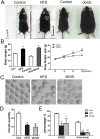
Figure 1
Figure 1. Obesity effects on mouse body weight and oocyte maturation.
(A,B) The body weights of HFD and ob/ob mice were significantly greater than those of control mice. (C) HFD and ob/ob mice had dark, smaller oocytes. (D) The numbers of oocytes from both obesity groups were reduced. (E) The proportions of GVBD in oocytes and polar body extrusion rates were reduced for oocytes from HFD and ob/ob induced mice with obesity.
Figure 2
Figure 2. Obesity disrupts meiotic spindle morphology and alters oocyte polarization.
(A) Oocytes from both groups of mice with obesity had misaligned chromosomes and disrupted spindle morphologies and the rates of abnormal spindle morphology were higher than that of control oocytes. (B) Actin fluorescence intensity at the plasma membrane and in the cytoplasm was lower than that in control MI oocytes. (C) CGs were uniformly localized at the plasma membrane and CG signals were weaker in oocytes from mice with obesity.
Figure 3
Figure 3. Obesity results in oocyte oxidative stress.
(A) DHE expression was increased in oocytes from both obesity groups as were the fluorescence intensity ratios for oxidative stress in these oocytes. (B) ROS levels determined by fluorescent staining and the fluorescence intensity ratios of ROS. (C) Fold-changes in oxidative stress related genes’ mRNA expression were altered in oocytes from both HFD and ob/ob induced mice with obesity.
Figure 4
Figure 4. Obesity disrupts distributions and morphology of mitochondria and induces early oocyte apoptosis.
(A) Mitochondria distribution patterns in oocytes at the GV and MI stages. The distribution patterns of mitochondria in oocytes from HFD and ob/ob mice were primarily clustered. (B) Transmission electron microscopy images showing mitochondrial morphologies in oocytes from HFD and control mice. Arrows indicate abnormal mitochondria that lack cristae and have broken membranes. (C) Early apoptosis signals in oocytes were assessed using Annexin V/PI kits. Oocytes from HFD and ob/ob mice had bright fluorescent signals. (D) Bak and Bcl-2 mRNA expressions in oocytes. Both obesity groups had higher fold-changes in mRNA expressions.
Figure 5
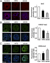
Figure 5. Obesity affects epigenetic modifications in oocytes.
(A) 5 mC genomic contents. 5 mC expression levels in oocytes from HFD and ob/ob mice with obesity were reduced compared to that in controls. (B) H3K27-me2 genomic contents. The H3K27-me2 fluorescence intensity ratio was reduced in oocytes from both HFD and ob/ob induced mice with obesity as compared to that in controls. (C) H3K9-me2 genomic contents. The H3K9-me2 fluorescence intensity ratio was increased in oocytes from both HFD and ob/ob induced mice with obesity as compared to that in controls.

References

    1. Penzias A. S. Recurrent IVF failure: other factors. Fertil Steril 97, 1033–1038 (2012). - PubMed
    1. Chu S. Y. et al. Maternal obesity and risk of stillbirth: a metaanalysis. Am J Obstet Gynecol 197, 223–228 (2007). - PubMed
    1. Samuelsson A. M. et al. Diet-Induced Obesity in Female Mice Leads to Offspring Hyperphagia, Adiposity, Hypertension, and Insulin Resistance: A Novel Murine Model of Developmental Programming. Hypertension 51, 383–392 (2008). - PubMed
    1. Robker R. L. Evidence that obesity alters the quality of oocytes and embryos. Pathophysiology 15, 115–121 (2008). - PubMed
    1. Jungheim E. S. et al. Associations between free fatty acids, cumulus oocyte complex morphology and ovarian function during in vitro fertilization. Fertil Steril 95, 1970–1974 (2011). - PMC - PubMed

Publication types

MeSH terms