. 2015 Dec 24:6:629.
doi: 10.3389/fimmu.2015.00629. eCollection 2015.

Systemic Inflammation in Cachexia - Is Tumor Cytokine Expression Profile the Culprit?

Affiliations

Systemic Inflammation in Cachexia - Is Tumor Cytokine Expression Profile the Culprit?

Emidio M de Matos-Neto et al. Front Immunol. .

Abstract

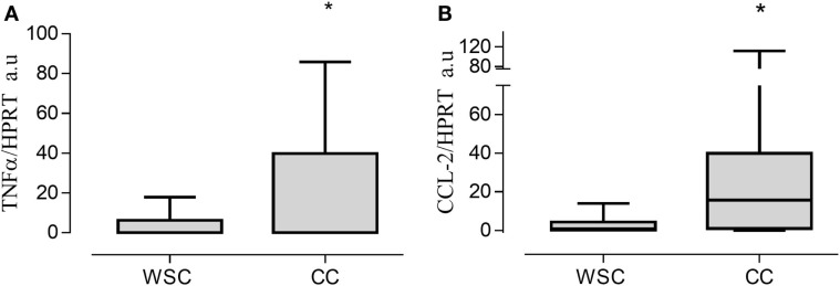
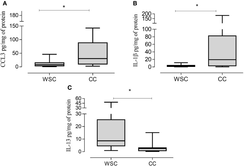
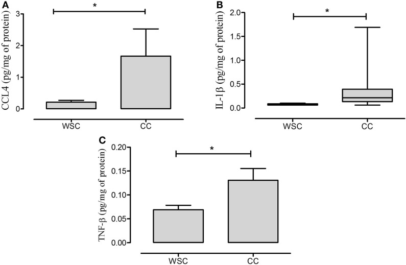
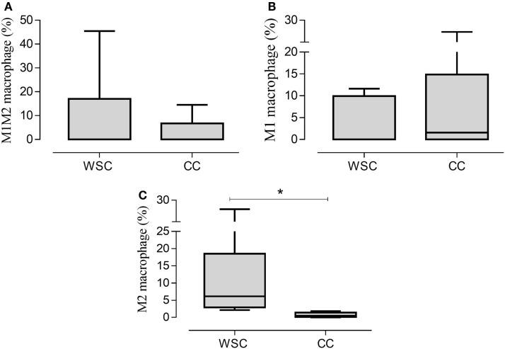
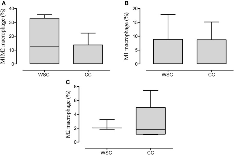
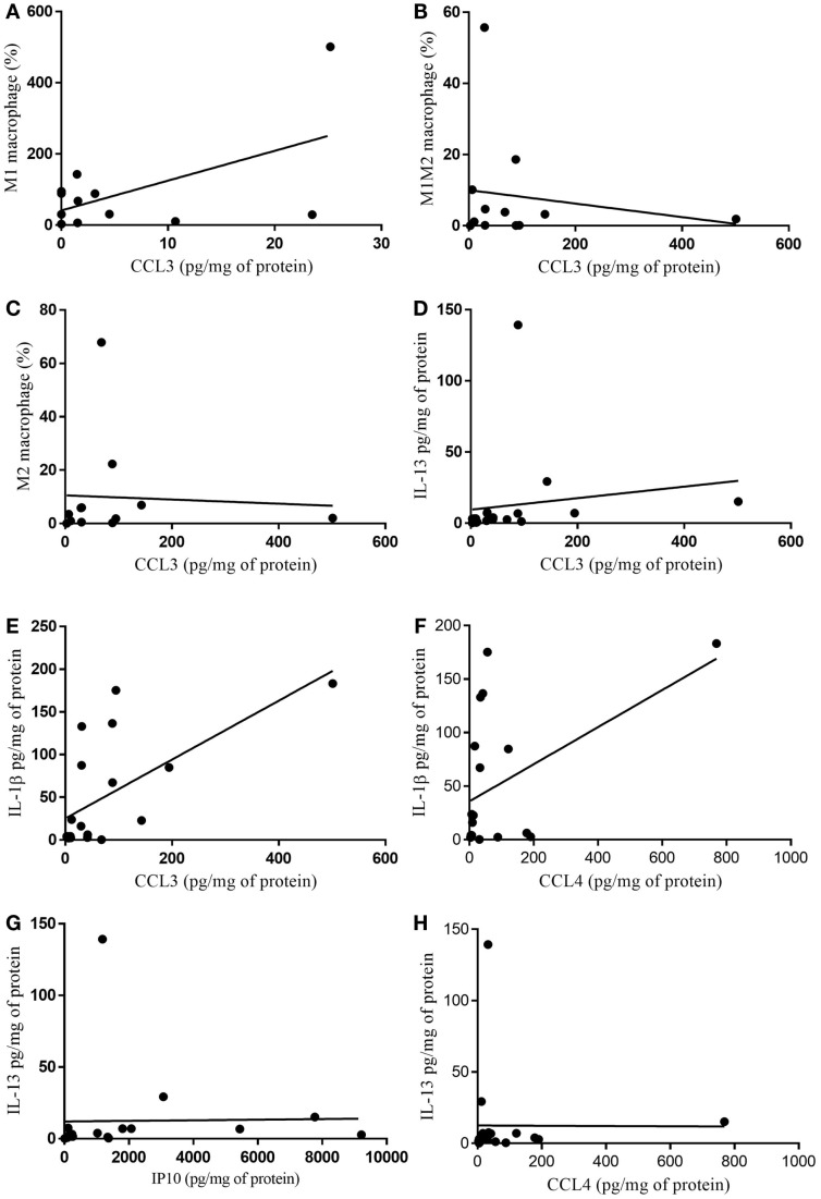
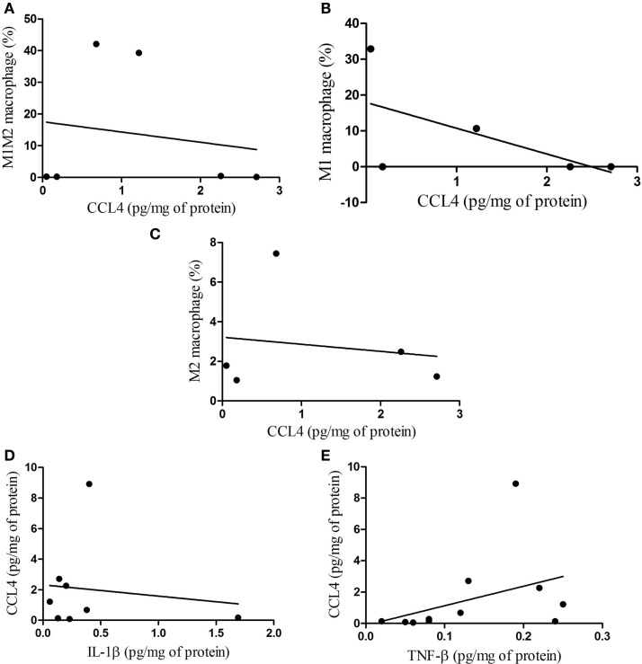
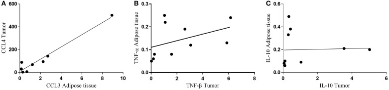
Cachexia affects about 80% of gastrointestinal cancer patients. This multifactorial syndrome resulting in involuntary and continuous weight loss is accompanied by systemic inflammation and immune cell infiltration in various tissues. Understanding the interactions among tumor, immune cells, and peripheral tissues could help attenuating systemic inflammation. Therefore, we investigated inflammation in the subcutaneous adipose tissue and in the tumor, in weight stable and cachectic cancer patients with same diagnosis, in order to establish correlations between tumor microenvironment and secretory pattern with adipose tissue and systemic inflammation. Infiltrating monocyte phenotypes of subcutaneous and tumor vascular-stromal fraction were identified by flow cytometry. Gene and protein expression of inflammatory and chemotactic factors was measured with qRT-PCR and Multiplex Magpix(®) system, respectively. Subcutaneous vascular-stromal fraction exhibited no differences in regard to macrophage subtypes, while in the tumor, the percentage of M2 macrophages was decreased in the cachectic patients, in comparison to weight-stable counterparts. CCL3, CCL4, and IL-1β expression was higher in the adipose tissue and tumor tissue in the cachectic group. In both tissues, chemotactic factors were positively correlated with IL-1β. Furthermore, positive correlations were found for the content of chemoattractants and cytokines in the tumor and adipose tissue. The results strongly suggest that the crosstalk between the tumor and peripheral tissues is more pronounced in cachectic patients, compared to weight-stable patients with the same tumor diagnosis.

Keywords: cancer cachexia; inflammatory cells; tumor-adipose tissue crosstalk macrophages.


Figures

Figure 1
Figure 1
Gene expression in tumor tissue. Data expressed as mean ± SE or as median [first quartile; third quartile]. *Significant difference between WSC vs. CC. Expression of target genes was normalized to the reference HPRT1. TNF-α, tumor necrosis factor α (A); CCL2, chemokine (C–C motif) ligand 2 (B); Arbitrary units, AU. WSC (n = 10) and CC (n = 14).
Figure 2
Figure 2
CCL3, IL-1β, and IL-13 protein expression in tumor samples. Data expressed as median [first quartile; third quartile]. *Significant difference between WSC vs. CC. CCL3, chemokine (C–C motif) ligand 3 (A); IL-1β, interleukin 1β (B); IL-13, interleukin 13 (C). WSC (n = 11) and CC (n = 12).
Figure 3
Figure 3
CCL4, IL-1β, and TNF-β protein expression in subcutaneous adipose tissue. Data expressed as mean ± SE. *Significant difference CC vs. WSC group. CCL4, chemokine (C–C motif) ligand 4 (A); IL-1β, interleukin 1β (B); TNF-β, tumor necrosis factor β (C). WSC (n = 11) and CC (n = 12).
Figure 4
Figure 4
Percentage of the phenotypes of macrophage populations in the tumor microenvironment. Data expressed as median [first quartile; third quartile] or median ± SE. *Significant difference between WSC and CC. Tumor samples WSC and CC (n = 5). M1M2 macrophage (A); M1 macrophage (B); M2 macrophage (C).
Figure 5
Figure 5
Percentage of the phenotypes of macrophage in subcutaneous adipose tissue. Data expressed as median [first quartile; third quartile]. Stromal-vascular fraction of subcutaneous adipose tissue: WSC (n = 4) and CC (n = 5). M1M2 macrophage (A); M1 macrophage (B); M2 macrophage (C).
Figure 6
Figure 6
Correlation of cytokine protein expression and % of infiltrating immune cells in tumor. (A) CCL3/M1 macrophage (%) p = 0.938; (B) CCL3/M1M2 macrophage (%) p = 0.956; (C) CCL3/M2 macrophage (%) p = 0.342; (D) CCL3/IL-13 p = 0.0089; (E) CCL3/IL-1β p = 0.0059; (F) CCL4/IL-1β p = 0.089; (G) IP10/IL-13 p = 0.057; (H) CCL4/IL-13 p = 0.147.
Figure 7
Figure 7
Correlations between macrophage phenotypes and CCL4 protein, and between CCL4 and IL-1β, TNF-β in subcutaneous adipose tissue. (A) M1M2/CCL4, p = 0.787; (B) M1/CCL4, p = 0.321; (C) M2/CCL4, p = 0.790 and correlations between CCL4 protein and IL-1β, TNF-β (D) CCL4/IL-1β, p = 0.955; (E) CCL4/TNF-β, p = 0.041.
Figure 8
Figure 8
Correlation between protein expression of inflammatory factors in subcutaneous adipose tissue and tumor. (A) CCL4 tumor/CCL3 adipose tissue; (B) TNF-α adipose tissue/TNF-β tumor; (C) IL-10 adipose tissue/IL-10 tumor.

References

    1. Fearon K, Strasser F, Anker SD, Bosaeus I, Bruera E, Fainsinger RL, et al. Definition and classification of cancer cachexia: an international consensus. Lancet Oncol (2011) 12:489–95.10.1016/S1470-2045(10)70218-7 - DOI - PubMed
    1. Argilés JM, Busquets S, Stemmler B, López-Soriano FJ. Cachexia and sarcopenia: mechanisms and potential targets for intervention. Curr Opin Pharmacol (2015) 22:100–6.10.1016/j.coph.2015.04.003 - DOI - PubMed
    1. Fearon K, Arends J, Baracos V. Understanding the mechanisms and treatment options in cancer cachexia. Nat Rev Clin Oncol (2013) 10:90–9.10.1038/nrclinonc.2012.209 - DOI - PubMed
    1. Khan S, Tisdale MJ. Catabolism of adipose tissue by a tumour-produced lipid-mobilising factor. Int J Cancer (1999) 80:444–7.10.1002/(SICI)1097-0215(19990129)80:3<444::AID-IJC18>3.0.CO;2-U - DOI - PubMed
    1. Todorov P, Cariuk P, Mcdevitt T, Coles B, Fearon K, Tisdale M. Characterization of a cancer cachectic factor. Nature (1996) 379:739–42.10.1038/379739a0 - DOI - PubMed

LinkOut - more resources