. 2016 Feb;47(2):498-504.
doi: 10.1161/STROKEAHA.115.012079. Epub 2016 Jan 5.

Interleukin-4 Is Essential for Microglia/Macrophage M2 Polarization and Long-Term Recovery After Cerebral Ischemia

Affiliations

Interleukin-4 Is Essential for Microglia/Macrophage M2 Polarization and Long-Term Recovery After Cerebral Ischemia

Xiangrong Liu et al. Stroke. 2016 Feb.

Abstract

Background and purpose: Interleukin-4 (IL-4) is a unique cytokine that may contribute to brain repair by regulating microglia/macrophage functions. Thus, we examined the effect of IL-4 on long-term recovery and microglia/macrophage polarization in 2 well-established stroke models.

Methods: Transient middle cerebral artery occlusion or permanent distal middle cerebral artery occlusion was induced in wild-type and IL-4 knockout C57/BL6 mice. In a separate cohort of wild-type animals, IL-4 (60 ng/d for 7 days) or vehicle was infused into the cerebroventricle after transient middle cerebral artery occlusion. Behavioral outcomes were assessed by the Rotarod, corner, foot fault, and Morris water maze tests. Neuronal tissue loss was verified by 2 independent neuron markers. Markers of classically activated (M1) and alternatively activated (M2) microglia were assessed by real-time polymerase chain reaction, immunofluorescence, and flow cytometry.

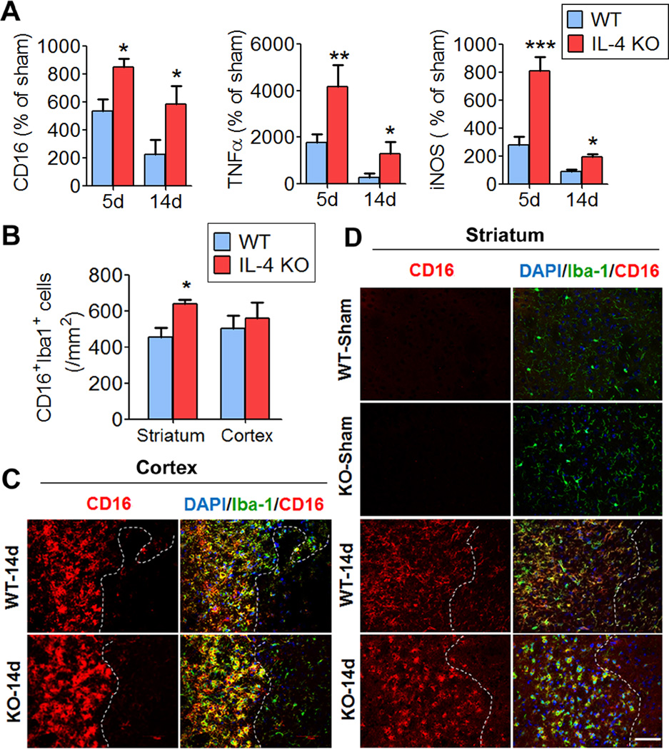
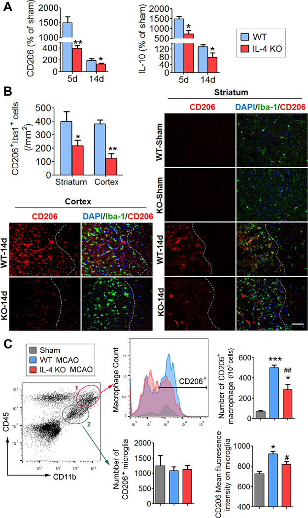
Results: Loss of IL-4 exacerbated sensorimotor deficits and impaired cognitive functions ≤21 days post injury. In contrast to the delayed deterioration of neurological functions, IL-4 deficiency increased neuronal tissue loss only in the acute phase (5 days) after stroke and had no impact on neuronal tissue loss 14 or 21 days post injury. Loss of IL-4 promoted expression of M1 microglia/macrophage markers and impaired expression of M2 markers at 5 and 14 days post injury. Administration of IL-4 into the ischemic brain also enhanced long-term functional recovery.

Conclusions: The cytokine IL-4 improves long-term neurological outcomes after stroke, perhaps through M2 phenotype induction in microglia/macrophages. These results are the first to suggest that immunomodulation with IL-4 is a promising approach to promote long-term functional recovery after stroke.

Keywords: flow cytometry; interleukin-4; macrophages; neurological function; stroke.


Figures

Figure 1
Figure 1. Loss of IL-4 exacerbates long-term sensorimotor dysfunction after ischemia
A–B, RNA extracts were prepared from ischemic brains at 1, 3, 5, 7, and 14 days after tMCAO from wild type (WT) or IL-4 KO mice. The mRNA levels of IL-4 (A) and IL-4 receptor α (B) were detected by real time PCR. *p≤0.05, **p≤0.01, vs WT sham mice. C, Left: representative images of cerebral blood flow (CBF) before cerebral ischemia, 5 minutes after ischemia, and 5 minutes after reperfusion. Right, Quantification of CBF. Results are expressed as percent change from baseline (pre-ischemia). IL-4 KO mice showed similar CBF compared with WT mice. n=6 to 8 mice per group. D, Sensorimotor function was evaluated up to 14d after tMCAO or sham operation by the Rotarod (left) and corner tests (right) in IL-4 KO and WT mice. n=9 for WT, n=12 for IL-4 KO. Shown are the mean ± SEM. *p≤0.05, **p≤0.01 vs WT.
Figure 2
Figure 2. Loss of IL-4 exacerbates cognitive deficits after cerebral ischemia
Long-term cognitive functions were assessed by the Morris water maze. A, The typical swim path of wild type (WT) or IL-4 KO mice in the Morris water maze test. B, Time to reach the submerged platform in the water maze 17 to 20 days after ischemia. C, Spatial memory of the location of the previously submerged platform 21 days after ischemia. Spatial memory is expressed as the time spent in the goal quadrant when the platform was removed. D. Average swim speed for mice in the Morris water maze. n=9 for WT, n=12 for IL-4 KO. All data are presented as mean ± SEM, *p≤0.05, **p≤0.01, ***p≤0.001.
Figure 3
Figure 3. Loss of IL-4 increases brain tissue loss at early but not late stages after ischemia
A, Representative brain slices with MAP2-negative infarcts in wild-type mice and IL-4 KO mice at 5 d, 14 d, and 21 d after ischemia. B–D, Quantification of infarct size by loss of MAP2 immunostaining in the cortex (B), striatum (C), and total brain (D). n=6 mice/group. Data were expressed as mean ± SEM. *, p≤0 .05; ** p≤0 .01 compared to WT mice.
Figure 4
Figure 4. Loss of IL-4 promotes the M1 phenotype in microglia/macrophages after ischemic stroke
A, M1 marker (CD16, TNFα and iNOS) mRNA levels. RT-PCR was performed using total RNA extracted from ischemic brains at 5 and 14 days after tMCAO or from sham-operated brains. Data are expressed as % of sham-operated controls. n=4–6 per group. B, Quantification of the number of CD16+ microglia/macrophages at the inner boundary of the infarct in wild-type and IL-4 KO mice at 14 d after tMCAO or sham-operation. n=4–5 mice/group. Data were expressed as mean ± SEM. *p≤0.05, **p≤0.01, ***p≤0.001 vs WT. C–D, Representative dual immunofluorescent staining of CD16 and Iba1 in the cortex (C) and striatum (D) in brain sections obtained from ischemic mice at 14 days after MCAO. Scale bar: 50 µm.
Figure 5
Figure 5. Loss of IL-4 reduces expression of the M2 phenotype in microglia/macrophages after ischemic stroke
A, M2 marker (CD206 and IL-10) mRNA levels. RT-PCR was performed using total RNA extracted from ischemic brains at 5 and 14 days after tMCAO or from sham-operated brains. Data are expressed as % of sham-operated controls. n=4–6 per group. B, Quantification of the number of CD206+ microglia/macrophage at the inner boundary of the infarct in wild-type and IL-4 KO mice 14 d after tMCAO or sham operation. n=4–5 mice/group. Data were expressed as mean ± SEM. *p≤0.05, **p≤0.01 vs WT. Images show representative double immunofluorescent staining of CD206 and Iba1 in the cortex and striatum in brain sections obtained from ischemic mice. Scale bar: 50 µm. C, Flow cytometry analyses of CD206+ M2 macrophages (CD45highCD11b+) and microglia (CD45intermediateCD11b+) in the ischemic brain in wild-type and IL-4 KO mice. n=3–5 mice/group. Data were expressed as mean ± SEM. *p≤0.05, ***p≤0.001 vs WT sham. #p≤0.05, ##p≤0.01 vs WT MCAO.
Figure 6
Figure 6. Ventricular administration of IL-4 after ischemia improves long-term neurological functions
Mice received icv infusions of IL-4 (60 ng/d) or vehicle 6h to 7d after tMCAO. A–B, Sensorimotor functions were assessed up to 35d after tMCAO or sham operation by the Rotarod (A) and foot fault (B) tests. C, Long-term cognitive functions were evaluated in the Morris water maze. IL-4-treated mice showed improvements in cognitive function, as reflected in shorter escape times (spatial learning) and extended time spent in the target quadrant (spatial memory), with no change in swim speed. D, Brain tissue loss was measured on NeuN-stained coronal sections 14d and 35d after tMCAO. Representative images show NeuN immunostaining at 14d after tMCAO. n=6 for sham, n=9 for WT, n=13 for IL-4 KO. Shown are the mean±SEM. *p≤0.05, **p≤0.01 IL-4 vs. PBS or as indicated.

References

    1. Miron VE, Boyd A, Zhao JW, Yuen TJ, Ruckh JM, Shadrach JL, et al. M2 microglia and macrophages drive oligodendrocyte differentiation during cns remyelination. Nature neuroscience. 2013;16:1211–1218. - PMC - PubMed
    1. Hu X, Li P, Guo Y, Wang H, Leak RK, Chen S, et al. Microglia/macrophage polarization dynamics reveal novel mechanism of injury expansion after focal cerebral ischemia. Stroke; a journal of cerebral circulation. 2012;43:3063–3070. - PubMed
    1. Wang G, Zhang J, Hu X, Zhang L, Mao L, Jiang X, et al. Microglia/macrophage polarization dynamics in white matter after traumatic brain injury. Journal of cerebral blood flow and metabolism : official journal of the International Society of Cerebral Blood Flow and Metabolism. 2013;33:1864–1874. - PMC - PubMed
    1. Butovsky O, Ziv Y, Schwartz A, Landa G, Talpalar AE, Pluchino S, et al. Microglia activated by il-4 or ifn-gamma differentially induce neurogenesis and oligodendrogenesis from adult stem/progenitor cells. Molecular and cellular neurosciences. 2006;31:149–160. - PubMed
    1. Kotter MR, Zhao C, van Rooijen N, Franklin RJ. Macrophage-depletion induced impairment of experimental cns remyelination is associated with a reduced oligodendrocyte progenitor cell response and altered growth factor expression. Neurobiology of disease. 2005;18:166–175. - PubMed

Publication types

MeSH terms