Clinical Trial
. 2016 Sep;9(5):1163-1172.
doi: 10.1038/mi.2015.136. Epub 2016 Jan 6.

Characterization of the human immune cell network at the gingival barrier

Affiliations
Clinical Trial

Characterization of the human immune cell network at the gingival barrier

Nicolas Dutzan et al. Mucosal Immunol. 2016 Sep.

Abstract

The oral mucosa is a barrier site constantly exposed to rich and diverse commensal microbial communities, yet little is known of the immune cell network maintaining immune homeostasis at this interface. We have performed a detailed characterization of the immune cell subsets of the oral cavity in a large cohort of healthy subjects. We focused our characterization on the gingival interface, a particularly vulnerable mucosal site, with thin epithelial lining and constant exposure to the tooth adherent biofilm. In health, we find a predominance of T cells, minimal B cells, a large presence of granulocytes/neutrophils, a sophisticated network of professional antigen-presenting cells (APCs), and a small population of innate lymphoid cells (ILCs) policing the gingival barrier. We further characterize cellular subtypes in health and interrogate shifts in immune cell populations in the common oral inflammatory disease periodontitis. In disease, we document an increase in neutrophils and an upregulation of interleukin-17 (IL-17) responses. We identify the main source of IL-17 in health and Periodontitis within the CD4(+) T-cell compartment. Collectively, our studies provide a first view of the landscape of physiologic oral immunity and serve as a baseline for the characterization of local immunopathology.


Figures

Figure 1
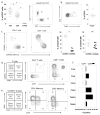
Figure 1. Major immune cell populations in oral mucosal tissues
Oral biopsies (4mm) were harvested from buccal mucosa and gingiva of healthy patients. (a-b) H&E staining of buccal (a) and gingival biopsies (bii) shown. Schematic depicting gingival area (Bi). (c) Percentage of hematopoietic (CD45+) cells per biopsy type (n=5, *p<0.05). (d-g) Characterization of the major hematopoietic cell populations in buccal oral mucosa and gingiva in health Plots of SSC and HLA-DR expression allowed separation of Lymphocytes (Lymph), granulocytes (Gran) and dendritic cells-monocytes-macrophages (DC-Mac) subsets. Granulocytes stained for CD15 and CD117, Lymphocytes stained for CD3 and CD19 (D=Buccal, E=Gingiva, major percentages shown). (f) Percentages of Lymph, DC-Mac and Gran in buccal mucosa and gingiva within the CD45+ compartment (n=5, *p<0.05). (g) Percentages of Lymph, DC-Mac and Gran in gingiva within the CD45+ compartment (n=15, *p<0.05).
Figure 2
Figure 2. The antigen presenting cell network in human gingiva in health
Analysis of DC-Mac cell subsets by flow cytometry (a) Cells gated from Single/Live/CD45+/SCCmid/hi/HLA DR+/Lineage (CD3/CD19/CD20), were evaluated for auto-fluorescence and auto fluorescent positive cells were stained for CD14. (b) Frequency of resident macrophages (AF+CD14+), recruited monocytes (AFCD14+) and DC subsets (AFCD14) in human gingiva (n=10. *p<0.05) (c) Single/Live/CD45+/SCCmid/hi/HLA-DR+/Lineage/AF (CD3/CD19/CD20) were stained for CD1a and EpCAM expression. (d) Frequency of CD1a (Langerhans cells), CD1c and CD141 dendritic cell subsets in healthy human gingival tissues (n=10, *p<0.05).(e) Single/Live/CD45+/SCCmid/hi/HLA-DR+/Lineage(CD3/CD19/CD20) were evaluated for CD14, CD1c, CD141, CD11c, CD1c.
Figure 3
Figure 3. T cell network in human gingiva in health
(a) Major T cell subsets (CD4, CD8, TCRγδ) in healthy gingival tissues. Single/Live/CD45+/HLA-DR/SCClow/CD19/CD3+ cells and were analyzed for CD4, CD8 and TCRγδ markers, frequency and representative facs plot shown (n=25, *p<0.05). (b) CD3+CD4+ cells evaluated for Foxp3 expression, frequency and representative plot shown (n=13). (c) Evaluation of CD45RO and CD45RA within the CD4 and CD8 compartment. Representative plots and frequencies shown (n=8) (d-e). Naïve and Memory T cell subsets in gingival tissues in health. (d). Single/Live/CD45+/HLA-DR/SCClow/CD19/CD3+ were evaluated for expression of CD45RA and CCR7 within the CD4 and CD8 compartments. Representative plots and frequency of populations shown (n=5). (e) Single/Live/CD45+/HLA-DR/SCClow/CD19/CD3+/CD45RO+ were analyzed for the expression for CD69 and CCR7 within the CD4 and CD8 compartments. Representative plots shown and (f) and frequency of subpopulations populations shown (n=5, *p<0.05).
Figure 4
Figure 4. Cytokine profiles of T cell and ILC subsets
(a) Ex-vivo IFN-γ and IL-17A production by T cell subsets. Cells were stimulated using PMA/Ionomycin and frequencies of IFN/IL17 secreting cells was evaluated in CD4+,CD8+ and TCRγδ+ cells. Representative plots shown (n=10). (b) Single/Live/CD45+ were evaluated for presence of Lineage specific markers Lin= (CD3/CD19/CD20/CD1a/CD11c/CD14/FcεR1α/CD16/CD34) and Lin- cells were evaluated following stimulation for secretion of IFN/IL17 (representative plots shown, n=5). (c) Phenotypic analysis of the lineage negative population. Lin-cells were evaluated for expression of CD127 (ILC marker). Lin-CD127 were evaluated for CD56 and NKp46. Lin-CD127+ cells were evaluated for CD161+, CRTH2, NKp44, NKp46.
Figure 5
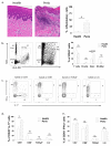
Figure 5
Shifts of major immune cell population in Chronic Periodontitis. (a) Histological sections (H&E) of Health (Health) and Chronic Periodontitis (Perio). Quantification of percentage of inflammatory cells in health and disease tissues (n=5 per group, 10- 20× fields counted per tissue). (b) Flow cytometric analysis of major immune cells in gingival tissues in health and in periodontitis. Cells were gated on Single/Live/CD45+ and analyzed according their internal granularity (SSC) and the expression of HLA-DR molecule. Granulocyte (Gran), DC-Mac and Lymphocyte (Lymph) gates marked also outlined. All cell subsets were confirmed with further flow cytometry for lineage markers. Frequencies of T (CD3) cells, B (CD19/20) cells, Granulocytes (CD15/CD16) and DC Mac (HLADR+CD19-) subpopulations were graphed. (c) Production of IL-17 and IFNγ in Periodontitis. Analysis of cytokine production within CD4+, CD8+ and Lin- cells (Lin=CD3/CD19). Representative plots shown (n=5 per group). (d-e) Graphs showing percentage of CD45+IL-17 producing cells (d) and (e) CD45+IFNγ producing cells within the CD4, CD8, TCRγδ and Lin- populations (n=5 per group, *p<0.05).

References

    1. Belkaid Y, Artis D. Immunity at the barriers. European journal of immunology. 2013;43:3096–3097. - PMC - PubMed
    1. Winning TA, Townsend GC. Oral mucosal embryology and histology. Clin Dermatol. 2000;18:499–511. - PubMed
    1. Costello EK, et al. Bacterial community variation in human body habitats across space and time. Science. 2009;326:1694–1697. - PMC - PubMed
    1. Human Microbiome Project, C A framework for human microbiome research. Nature. 2012;486:215–221. - PMC - PubMed
    1. Aas JA, Paster BJ, Stokes LN, Olsen I, Dewhirst FE. Defining the normal bacterial flora of the oral cavity. J Clin Microbiol. 2005;43:5721–5732. - PMC - PubMed

Publication types

MeSH terms