. 2016 Jan 6:6:18945.
doi: 10.1038/srep18945.

Effects of calcitriol on random skin flap survival in rats

Affiliations

Effects of calcitriol on random skin flap survival in rats

Kai-liang Zhou et al. Sci Rep. .

Abstract

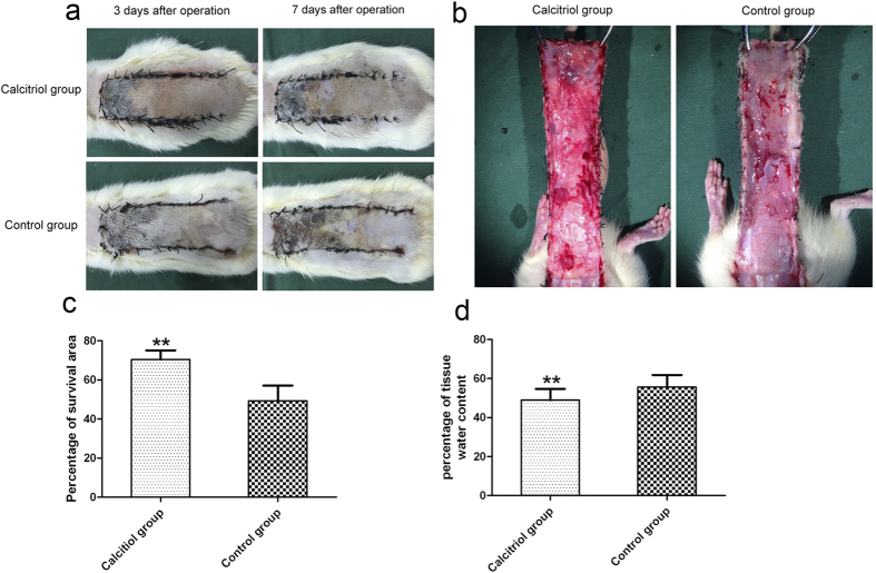
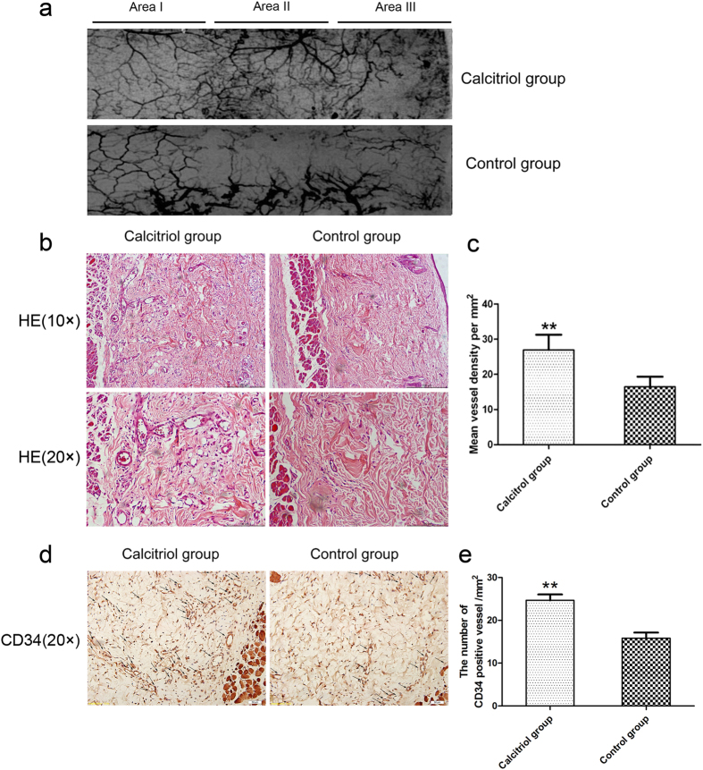
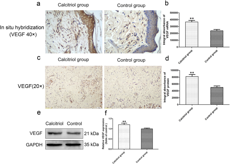
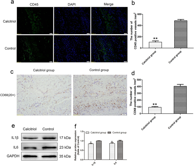
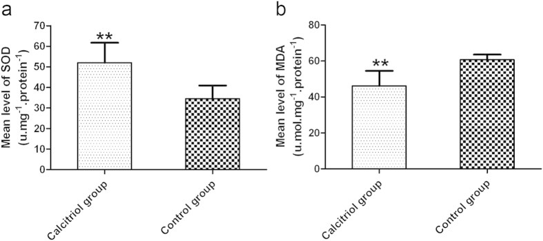
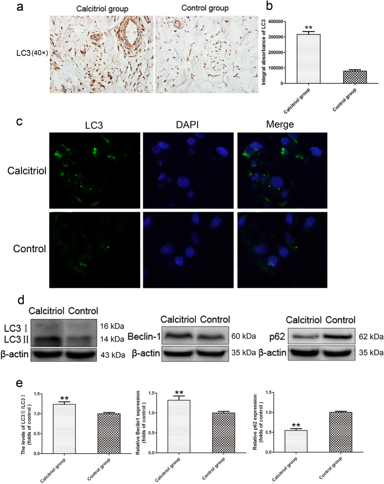
Calcitriol, a metabolite of vitamin D, is often used in osteoporosis clinics. However, the material has other bioactivities; for example, it accelerates angiogenesis, has anti-inflammatory properties, and inhibits oxidative stress. We investigated the effects of calcitriol in a random skin flap rat model. "McFarlane flap" models were established in 84 male Sprague Dawley rats, divided into two groups. One group received intraperitoneal injections of calcitriol (2 μg/kg/day) whereas control rats received intraperitoneal injections of saline. The percentage flap survival area and tissue water content were measured 7 days later, which showed that calcitriol improved flap survival area and reduced tissue edema. It also increased the mean vessel density and upregulated levels of VEGF mRNA/protein, both of which promote flap angiogenesis. Moreover, it decreased leukocyte and macrophage infiltration, reduced the inflammatory proteins IL1β and IL6, increased SOD activity, decreased MDA content, and upregulated the level of autophagy. Overall, our results suggest that calcitriol promotes skin flap survival by accelerating angiogenesis, having anti-inflammatory effects, reducing oxidative stress, and promoting autophagy.


Figures

Figure 1
Figure 1. Calcitriol improves flap survival area and reduces tissue edema.
(a) Digital photographs show the postoperative flaps of the calcitriol and control groups on Days 3 and 7. (b) Digital photographs show the tissue edema of postoperative flaps of each group on Day 7. (c) Histogram of percentages of survival area in the calcitriol group (70.42 ± 4.16%) and control group (49.20 ± 4.30%). (d) Histogram of percentages of tissue water content: 46.90 ± 5.45% in the calcitriol group and 57.45 ± 3.05% in the control group. Values are expressed as the mean ± SEM, n = 6 per group. **p <0.01, vs. control group.
Figure 2
Figure 2. Calcitriol promotes vascularization in skin flaps.
(a) Flap angiograms on postoperative Day 7 after surgery. (b) Neovascularization in calcitriol and control groups by H&E staining (original magnification ×100 and ×200). (c) Histogram of percentages of MVDs: calcitriol group (26.96 ± 4.33/mm2) and control group (16.48 ± 2.87/mm2). (d) CD34-positive vessels in the calcitriol and control groups as assessed by immunohistochemistry (original magnification ×200). (e) The numbers of CD34-positive vessels/mm2 were 24.67 ± 3.89/mm2 in the calcitriol group and 15.83 ± 3.19/mm2 in the control group. Values are expressed as means ± SEM, n = 6 per group. **p <0.01, vs. control group.
Figure 3
Figure 3. Calcitriol increases levels of VEGF mRNA/protein in skin flaps.
(a) In situ hybridization for VEGF mRNA in the calcitriol and control groups (original magnification × 400). (b) The integral absorbance (IA) values for VEGF mRNA were 366624.00 ± 50300.32 in the calcitriol group and 238306.20 ± 43730.12 in the control group. (c) VEGF expression in each group as assessed by immunohistochemistry (original magnification × 200). (d) The IA values of VEGF protein were 82087.16 ± 12687.08 in the calcitriol group and 50490.62 ± 8883.89 in the control group. (e) Protein expression of VEGF in each group, as assessed by Western blot analysis. The gels have been run under the same experimental conditions, and cropped blots are used here. The full-length gel images are available in Supplementary Fig. 3e. (f) Densitometry results of VEGF protein expression in the two groups. Values are expressed as means ± SEMs, n = 6 per group. **p < 0.01, vs. control group.
Figure 4
Figure 4. Calcitriol suppresses inflammatory response in skin flaps.
(a) Leukocyte infiltration in the calcitriol and control groups as assessed by immunofluorescence for CD45 (original magnification × 200). (b) The numbers of CD45-positive cells/mm2 were 105.71 ± 48.02/mm2 in the calcitriol group and 486.67 ± 48.52/mm2 in the control group. (c) Macrophage infiltration in each group as assessed by immunohistochemistry staining for CD68 (original magnification ×200). (d) The numbers of CD68-positive cells/mm2 were 94.49 ± 22.86/mm2 in the calcitriol group and 405.71 ± 59.35/mm2 in the control group. (e) Protein expression levels of IL1β and IL6 in each group as assessed by Western blot analysis. The gels have been run under the same experimental conditions, and cropped blots are used here. The full-length gel images are available in Supplementary Fig. 4e. (f) Optical densities of IL1β and IL6 proteins. Values are expressed as means ± SEMs, n = 6 per group. **p < 0.01, vs. control group, *p < 0.05, vs. control group.
Figure 5
Figure 5. Calcitriol attenuates oxidative stress in skin flaps.
(a) The levels of superoxide dismutase activity were 52.00 ± 9.76 U.mg−1protein−1 in the calcitriol group and 34.50 ± 6.44 U.mg−1protein−1 in the control. (b) Treatment with calcitriol resulted in a malondialdehyde content of 46.12 ± 8.33 nmol.mg−1protein−1 in the calcitriol group and 60.67 ± 2.88 nmol.mg−1protein−1 in the control group. Values are expressed as means ± SEMs, n = 6 per group. **p < 0.01, vs. control group.
Figure 6
Figure 6. Calcitriol upregulates autophagy in skin flaps.
(a) LC3II punctate dots were seen in the calcitriol and control groups in immunohistochemistry assessments (original magnification ×400). (b) Integral absorbance (IA) values for LC3 were 317103.53 ± 45034.03 in the calcitriol group and 80303.13 ± 19882.24 in the control group. (c) Immunofluorescence for LC3II punctate dots in the calcitriol group and control group. (d) Protein expression of LC3, Beclin1, and p62 in each group as assessed by Western blot analysis. The gels have been run under the same experimental conditions, and cropped blots are used here. The full-length gel images are available in Supplementary Fig. 6d. (e) Optical density analysis of ratios of LC3II/LC3I, Beclin1, and p62 protein expression in the calcitriol and control groups. Values are expressed as means ± SEMs, n = 6 per group. **p < 0.01, vs. control group.

References

    1. Akhavani M. A., Sivakumar B., Paleolog E. M. & Kang N. Angiogenesis and plastic surgery. J Plast Reconstr Aesthet Surg 61, 1425–1437 (2008). - PubMed
    1. Yang M., Sheng L., Li H., Weng R. & Li Q. F. Improvement of the skin flap survival with the bone marrow-derived mononuclear cells transplantation in a rat model. Microsurgery 30, 275–281 (2010). - PubMed
    1. Kim H. J. et al. Anti-inflammatory effects of anthocyanins from black soybean seed coat on the keratinocytes and ischemia-reperfusion injury in rat skin flaps. Microsurgery 32, 563–570 (2012). - PubMed
    1. Lima L. P. et al. Electroacupuncture attenuates oxidative stress in random skin flaps in rats. Aesthetic Plast Surg 36, 1230–1235 (2012). - PubMed
    1. Cardus A. et al. 1,25-dihydroxyvitamin D3 regulates VEGF production through a vitamin D response element in the VEGF promoter. Atherosclerosis 204, 85–89 (2009). - PubMed

Publication types

MeSH terms