. 2016 Jan 5;7(1):e01451-15.
doi: 10.1128/mBio.01451-15.

Characterization of a Pathogenic Full-Length cDNA Clone and Transmission Model for Porcine Epidemic Diarrhea Virus Strain PC22A

Affiliations

Characterization of a Pathogenic Full-Length cDNA Clone and Transmission Model for Porcine Epidemic Diarrhea Virus Strain PC22A

Anne Beall et al. mBio. .

Abstract

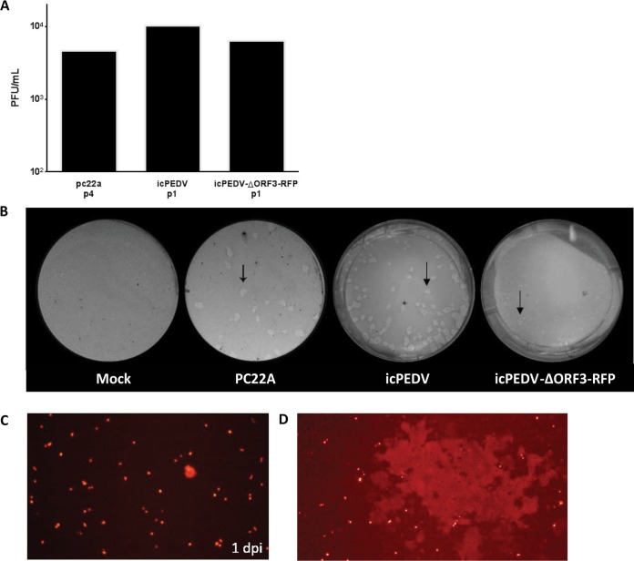
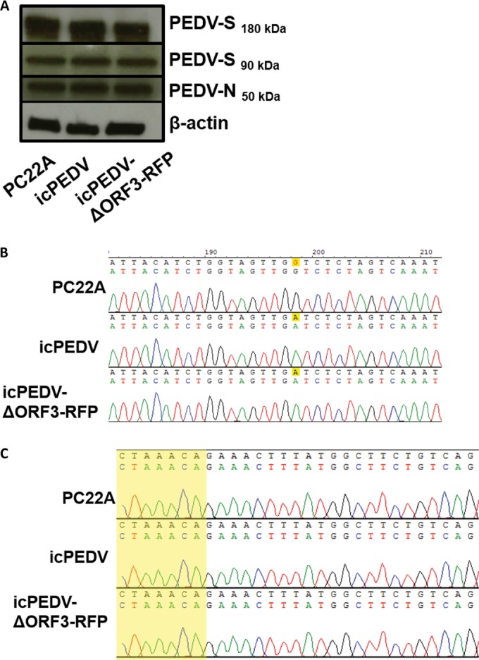
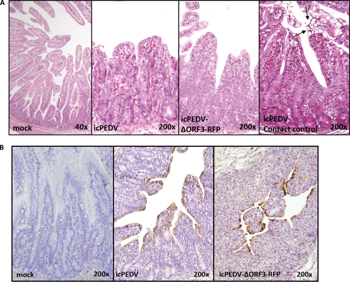
Porcine epidemic diarrhea virus (PEDV) is a highly pathogenic alphacoronavirus. In the United States, highly virulent PEDV strains cause between 80 and 100% mortality in suckling piglets and are rapidly transmitted between animals and farms. To study the genetic factors that regulate pathogenesis and transmission, we developed a molecular clone of PEDV strain PC22A. The infectious-clone-derived PEDV (icPEDV) replicated as efficiently as the parental virus in cell culture and in pigs, resulting in lethal disease in vivo. Importantly, recombinant PEDV was rapidly transmitted to uninoculated pigs via indirect contact, demonstrating virulence and efficient transmission while replicating phenotypes seen in the wild-type virus. Using reverse genetics, we removed open reading frame 3 (ORF3) and replaced this region with a red fluorescent protein (RFP) gene to generate icPEDV-ΔORF3-RFP. icPEDV-ΔORF3-RFP replicated efficiently in vitro and in vivo, was efficiently transmitted among pigs, and produced lethal disease outcomes. However, the diarrheic scores in icPEDV-ΔORF3-RFP-infected pigs were lower than those in wild-type-virus- or icPEDV-infected pigs, and the virus formed smaller plaques than those of PC22A. Together, these data describe the development of a robust reverse-genetics platform for identifying genetic factors that regulate pathogenic outcomes and transmission efficiency in vivo, providing key infrastructural developments for developing and evaluating the efficacy of live attenuated vaccines and therapeutics in a clinical setting.

Importance: Porcine epidemic diarrhea virus (PEDV) emerged in the United States in 2013 and has since killed 10% of U.S. farm pigs. Though the disease has been circulating internationally for decades, the lack of a rapid reverse-genetics platform for manipulating PEDV and identifying genetic factors that impact transmission and virulence has hindered the study of this important agricultural disease. Here, we present a DNA-based infectious-clone system that replicates the pathogenesis of circulating U.S. strain PC22A both in vitro and in piglets. This infectious clone can be used both to study the genetics, virulence, and transmission of PEDV coronavirus and to inform the creation of a live attenuated PEDV vaccine.


Figures

FIG 1
FIG 1
Schematic of the full-length PEDV genome and construction of the infectious PEDV cDNA clone and mutants. (A) PEDV genome, including ORF1a, ORF1b, and the spike (S), ORF3, envelope (E), membrane (M), and nucleocapsid (N) genes. (B) cDNA fragments comprising icPEDV. Restriction sites joining fragments are noted. (C) The BsaI site was removed from icPEDV, as indicated by the A to G nucleotide change in yellow. (D) PEDV-ΔORF3-RFP. Restriction sites were used to replace ORF3 with tomato-red and the ORF3 transcription regulatory sequence (TRS). 5′ and 3′ UTR, 5′ and 3′ untranslated regions.
FIG 2
FIG 2
Growth of icPEDV clones isolated from in vivo small intestine samples. (A) Titers of viral stocks isolated from small intestinal contents at the noted passages, harvested 2 dpi; (B) representative plaques (arrows) of mock-infected cells and cells infected with the parental PC22A strain, icPEDV, and icPEDV-ΔORF3-RFP, from left to right, at 2 dpi; (C) fluorescence microscopy of passage 0 icPEDV-ΔORF3-RFP in cell culture at 1 dpi; (D) syncytium formation of passage 0 icPEDV-ΔORF3-RFP.
FIG 3
FIG 3
Confirmatory studies of the infectious PEDV clone. (A) Western blot for PEDV spike (PEDV-S), before (180 kDa) and after (90 kDa) cleavage with trypsin, and nucleocapsid (PEDV-N). Protein was isolated from Vero cells infected with PC22A, icPEDV, or icPEDV-ΔORF3-RFP, which was harvested and sequenced 2 dpi. (B) An altered BsaI cloning site is conserved (yellow highlighting) after viral passage in piglets. (C) The transcription regulatory sequence (TRS) in the leader region of PEDV subgenomic transcripts is conserved between the wild type and icPEDV. The sequence is highlighted in yellow.
FIG 4
FIG 4
icPEDV mimics wild-type PEDV infection in gnotobiotic piglets. Gnotobiotic piglets were infected orally with 2 ml of the PEDV supernatant, and an uninoculated piglet was cohoused with them to determine transmission. All animals succumbed to illness or were euthanized due to illness at their final time points. (A) Mean RT-qPCR titers of fecal samples and fecal scores after viral inoculation. The line represents mean values, with individual piglet values shown by points. (B) RT-qPCR titers of fecal samples and fecal scores in cohoused transmission control piglets, days after contact with the inoculated piglets whose results are represented in panel A. The limit of detection in real-time (rt) RT-PCR figures (threshold cycle 37) is represented by a red line.
FIG 5
FIG 5
Histology and IHC staining of icPEDV-infected pig intestine. Gnotobiotic piglets were orally inoculated with 2 ml of PEDV. A contact control piglet was cohoused with an inoculated piglet(s) to determine transmission. All animals succumbed to illness or were euthanized due to illness at their final time points (PEDV, 1 dpi; icPEDV, 4 dpi; icPEDV-ΔORF3-RFP, 7 dpi; contact control, 4 dpi). All images show representative histological slides of jejunum specimens from mock-infected or infected animals. Histology of mock-, icPEDV-, or icPEDV-ORF3-RFP-infected animals, showing H&E staining and immunohistochemistry (IHC) staining (B) for PEDV nucleocapsid (N) protein using mouse anti-PEDV-N. A specimen from a contact piglet infected by icPEDV is also shown. Cell fusion and vacuolation were noted at the villus tips (arrows). IHC staining of icPEDV- or icPEDV-ΔORF3-RFP-infected animals whose results are represented in panel A is shown.

References

    1. Berry M, Gamieldien J, Fielding BC. 2015. Identification of new respiratory viruses in the new millennium. Viruses 7:996–1019. doi:10.3390/v7030996. - DOI - PMC - PubMed
    1. Stevenson GW, Hoang H, Schwartz KJ, Burrough ER, Sun D, Madson D, Cooper VL, Pillatzki A, Gauger P, Schmitt BJ, Koster LG, Killian ML, Yoon KJ. 2013. Emergence of porcine epidemic diarrhea virus in the United States: clinical signs, lesions, and viral genomic sequences. J Vet Diagn Invest 25:649–654. doi:10.1177/1040638713501675. - DOI - PubMed
    1. Madson DM, Magstadt DR, Arruda PHE, Hoang H, Sun D, Bower LP, Bhandari M, Burrough ER, Gauger PC, Pillatzki AE, Stevenson GW, Wilberts BL, Brodie J, Harmon KM, Wang C, Main RG, Zhang J, Yoon KJ. 2014. Pathogenesis of porcine epidemic diarrhea virus isolate (US/Iowa/18984/2013) in 3-week-old weaned pigs. Vet Microbiol 174:60–68. doi:10.1016/j.vetmic.2014.09.002. - DOI - PubMed
    1. Jung K, Annamalai T, Lu Z, Saif LJ. 2015. Comparative pathogenesis of US porcine epidemic diarrhea virus (PEDV) strain PC21A in conventional 9-day-old nursing piglets vs. 26-day-old weaned pigs. Vet Microbiol 178:31–40. doi:10.1016/j.vetmic.2015.04.022. - DOI - PMC - PubMed
    1. USDA-APHIS. 2015. Swine enteric coronavirus diseases (SECD), including porcine epidemic diarrhea virus (PEDv). USDA, Animal and Plant Health Inspection Service; https://www.aphis.usda.gov/wps/portal/aphis/ourfocus/animalhealth?urile=... Accessed 1 July 2015.

Publication types

MeSH terms

LinkOut - more resources