. 2016 Jan 5;7(1):e01865-15.
doi: 10.1128/mBio.01865-15.

Identification of Proteins Bound to Dengue Viral RNA In Vivo Reveals New Host Proteins Important for Virus Replication

Affiliations

Identification of Proteins Bound to Dengue Viral RNA In Vivo Reveals New Host Proteins Important for Virus Replication

Stacia L Phillips et al. mBio. .

Abstract

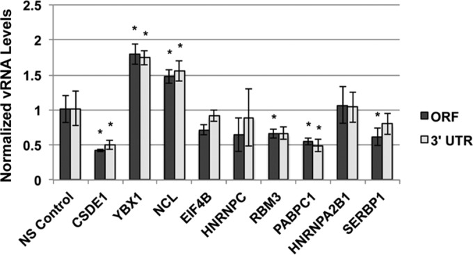
Dengue virus is the most prevalent cause of arthropod-borne infection worldwide. Due to the limited coding capacity of the viral genome and the complexity of the viral life cycle, host cell proteins play essential roles throughout the course of viral infection. Host RNA-binding proteins mediate various aspects of virus replication through their physical interactions with viral RNA. Here we describe a technique designed to identify such interactions in the context of infected cells using UV cross-linking followed by antisense-mediated affinity purification and mass spectrometry. Using this approach, we identified interactions, several of them novel, between host proteins and dengue viral RNA in infected Huh7 cells. Most of these interactions were subsequently validated using RNA immunoprecipitation. Using small interfering RNA (siRNA)-mediated gene silencing, we showed that more than half of these host proteins are likely involved in regulating virus replication, demonstrating the utility of this method in identifying biologically relevant interactions that may not be identified using traditional in vitro approaches.

Importance: Dengue virus is the most prevalent cause of arthropod-borne infection worldwide. Viral RNA molecules physically interact with cellular RNA-binding proteins (RBPs) throughout the course of infection; the identification of such interactions will lead to the elucidation of the molecular mechanisms of virus replication. Until now, the identification of host proteins bound to dengue viral RNA has been accomplished using in vitro strategies. Here, we used a method for the specific purification of dengue viral ribonucleoprotein (RNP) complexes from infected cells and subsequently identified the associated proteins by mass spectrometry. We then validated a functional role for the majority of these proteins in mediating efficient virus replication. This approach has broad relevance to virology and RNA biology, as it could theoretically be used to purify any viral RNP complex of interest.


Figures

FIG 1
FIG 1
Schematic of affinity purification technique. Cells were infected with dengue virus at an MOI of 1. Mock-infected cells served as a negative control. RNA-protein cross-links were induced 30 h postinfection by exposing the cells to 254 nm UV. Cells were lysed under denaturing conditions and incubated with biotinylated antisense DNA oligonucleotides. RNA-protein complexes were captured on streptavidin-coated magnetic beads. Proteins were liberated from the RNA by RNase treatment and identified by mass spectrometry.
FIG 2
FIG 2
Validation of RNA-protein interactions by RNA immunoprecipitation. Huh7 cells were infected with DENV2 NGC at an MOI of 1. Infected cell lysates were incubated with an isotype control antibody or an antibody directed against the indicated cellular proteins. Bound material was captured on Dynabeads protein G for analysis. (A) Western blot analysis of immunoprecipitated material. Input represents 10% of total input sample. IgG lanes represent 20% of immunoprecipitated protein. (B) RT-qPCR analysis of immunoprecipitated RNA. Statistical significance relative to DENV measured by Student’s t test: *, P ≤ 0.05; ◆, P ≤ 0.005; ●, P ≤ 0.0005.
FIG 3
FIG 3
High-content imaging of DENV infected cells. Huh7 cells were transfected with the indicated siRNAs and infected with DENV2 NGC at an MOI of 1 for 24 h. (A) Representative images used for high-content imaging. Nuclei are stained with Hoechst. Infected cells are indicated by the Alexa Fluor 488 signal from indirect immunofluorescent staining using a primary antibody recognizing the viral envelope protein. (B) Quantification of nuclei per condition by high-content imaging analysis. Data are representative of 4 independent experiments. Error bars represent standard errors of the mean (SEM) for three individual wells. (C) Percentage of cells infected measured by high-content imaging of cells expressing viral envelope protein. Statistical significance measured by Student’s t test: *, P ≤ 0.05; **, P ≤ 0.005; ***, P ≤ 0.0005.
FIG 4
FIG 4
Assessment of the role of host RBPs on vRNA levels. Huh7 cells were transfected with nonsilencing (NS) control siRNA or siRNA pools containing 4 unique siRNAs per target. Cells were infected with DENV 48 h posttransfection at an MOI of 1. Total cell-associated RNA was harvested 24 h postinfection, and levels of v RNA accumulation were measured by RT-qPCR using primers that amplify a region in the middle of the viral coding sequence (ORF) or in the 3′ UTR. Statistical significance measured by Student’s t test: *, P ≤ 0.05.
FIG 5
FIG 5
Measurement of viral titer after host RBP depletion. Huh7 cells were transfected with nonsilencing (NS) control siRNA or siRNA pools containing 4 unique siRNAs per target. Cells were infected with DENV 48 h posttransfection at an MOI of 1. Supernatants were collected 24 h postinfection, and viral titer was determined by focus formation assay. Statistical significance measured by Student’s t test: *, P ≤ 0.05; **, P ≤ 0.005.

References

    1. Bhatt S, Gething PW, Brady OJ, Messina JP, Farlow AW, Moyes CL, Drake JM, Brownstein JS, Hoen AG, Sankoh O, Myers MF, George DB, Jaenisch T, Wint GRW, Simmons CP, Scott TW, Farrar JJ, Hay SI. 2013. The global distribution and burden of dengue. Nature 496:504–507. doi:10.1038/nature12060. - DOI - PMC - PubMed
    1. Daffis S, Szretter KJ, Schriewer J, Li J, Youn S, Errett J, Lin T, Schneller S, Zust R, Dong H, Thiel V, Sen GC, Fensterl V, Klimstra WB, Pierson TC, Buller RM, Gale M, Shi P, Diamond MS. 2010. 2′-O methylation of the viral mRNA cap evades host restriction by IFIT family members. Nature 468:452–456. doi:10.1038/nature09489. - DOI - PMC - PubMed
    1. Bidet K, Garcia-Blanco M. 2014. Flaviviral RNAs: weapons and targets in the war between virus and host. Biochem J 462:215–230. doi:10.1042/BJ20140456. - DOI - PubMed
    1. Moon SL, Anderson JR, Kumagai Y, Wilusz CJ, Akira S, Khromykh AA, Wilusz J. 2012. A noncoding RNA produced by arthropod-borne flaviviruses inhibits the cellular exoribonuclease XRN1 and alters host mRNA stability. RNA 18:2029–2040. doi:10.1261/rna.034330.112. - DOI - PMC - PubMed
    1. Clarke BD, Roby JA, Slonchak A, Khromykh AA. 2015. Functional non-coding RNAs derived from the flavivirus 3′ untranslated region. Virus Res 206:53–61. doi:10.1016/j.virusres.2015.01.026. - DOI - PubMed

Publication types

LinkOut - more resources