. 2016 Jan 6:9:1.
doi: 10.1186/s13045-015-0229-y.

MicroRNA-26a suppresses epithelial-mesenchymal transition in human hepatocellular carcinoma by repressing enhancer of zeste homolog 2

Affiliations

MicroRNA-26a suppresses epithelial-mesenchymal transition in human hepatocellular carcinoma by repressing enhancer of zeste homolog 2

De-Ning Ma et al. J Hematol Oncol. .

Abstract

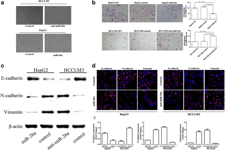
Background: Our previous study reported that microRNA-26a (miR-26a) inhibited tumor progression by inhibiting tumor angiogenesis and intratumoral macrophage infiltration in hepatocellular carcinoma (HCC). The direct roles of miR-26a on tumor cell invasion remain poorly understood. In this study, we aim to explore the mechanism of miR-26a in modulating epithelial-mesenchymal transition (EMT) in HCC.

Methods: In vitro cell morphology and cell migration were compared between the hepatoma cell lines HCCLM3 and HepG2, which were established in the previous study. Overexpression and down-regulation of miR-26a were induced in these cell lines, and Western blot and immunofluorescence assays were used to detect the expression of EMT markers. Xenograft nude mouse models were used to observe tumor growth and pulmonary metastasis. Immunohistochemical assays were conducted to study the relationships between miR-26a expression and enhancer of zeste homolog 2 (EZH2) and E-cadherin expression in human HCC samples.

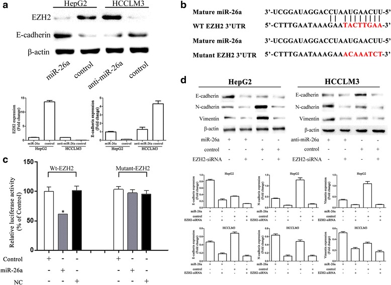
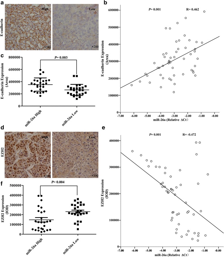
Results: Down-regulation of miR-26a in HCCLM3 and HepG2 cells resulted in an EMT-like cell morphology and high motility in vitro and increased in tumor growth and pulmonary metastasis in vivo. Through down-regulation of EZH2 expression and up-regulation of E-cadherin expression, miR-26a inhibited the EMT process in vitro and in vivo. Luciferase reporter assay showed that miR-26a directly interacted with EZH2 messenger RNA (mRNA). Furthermore, the expression of miR-26a was positively correlated with E-cadherin expression and inversely correlated with EZH2 expression in human HCC tissue.

Conclusions: miR-26a inhibited the EMT process in HCC by down-regulating EZH2 expression.


Figures

Fig. 1
Fig. 1
Down-regulation of miR-26a induced EMT in hepatoma cells in vitro. a Morphologic changes consistent with EMT (spindle-shaped cells with loss of polarity and increased intercellular separation) were observed in the HCCLM3-anti-miR-26a cells compared to control cells; HepG2 cells did not show morphological changes. b Cell migration through a Transwell chamber was compared between hepatoma cells transfected with miR-26a and control. The number of migrated cells was evaluated by counting 10 random fields at ×100 magnification. Western blot (c) and immunofluorescence assays (d) showed that down-regulation of miR-26a resulted in down-regulation of E-cadherin and up-regulation of N-cadherin and vimentin
Fig. 2
Fig. 2
EZH2 played a critical role in miR-26a modulating EMT process. a Western blot assay showed that miR-26a down-regulated EZH2 expression and up-regulated E-cadherin expression. b Diagram of the putative binding sequence of miR-26a in the 3′-UTR containing reporter constructs of EZH2 is shown. c Luciferase reporter assays showed that miR-26a significantly decreased the luciferase activity of wild-type EZH2 but not mutant EZH2. NC, the mock of precursor miRNA, *P < 0.05 compared with control. d EZH2 siRNA transfection in HCC cells eliminated the difference in E-cadherin miR-26 overexpression and down-regulation
Fig. 3
Fig. 3
miR-26a inhibited EMT process in vivo. a, b HCC subcutaneous tumor tissues (HCCLM3-control, HCCLM3-anti-miR-26a, HepG2-control, and HepG2-miR-26a) were implanted into nude mouse livers to establish the xenograft HCC models. Thirty-five days after implantation, the mice were euthanized, and their body weights and tumor volumes were assessed. c Quantification of bioluminescence of lung metastatic foci showed that down-regulation of miR-26a significantly accelerated pulmonary metastasis (arrows indicate metastatic foci in lung). HCCLM3-anti-miR-26a and HCCLM3-control tumors were compared by both bright field (b) and fluorescence (f) imaging. No pulmonary metastases occurred in HepG2-control xenografts and HepG2-miR-26a (up-regulation miR-26a) xenografts based on quantification of bioluminescence. d Immunohistochemistry revealed that E-cadherin expression was decreased and the expression level of N-cadherin and vimentin was increased in HCCLM3-anti-miR-26a tumors compared to HCCLM3-controls. In HepG2-miR-26a tumors, EMT markers were inhibited compared to HepG2-controls. In addition, EZH2 expression was inversely correlated with expression of E-cadherin in HCCLM3 and HepG2 tumors. Representative images are shown at ×200. *P < 0.05 compared to control
Fig. 4
Fig. 4
The relationships between miR-26a expression and the expression of EZH2 and E-cadherin in human HCC tissue. a A HCC tissue microarray was used to analyze E-cadherin expression using immunohistochemistry. Representative images are shown (×200). b miR-26a was positively associated with intratumoral E-cadherin expression (R = 0.462, P = 0.001). c Patients with high miR-26a expression had significantly higher E-cadherin expression compared with those with low miR-26a expression (P = 0.003). d Representative images of EZH2 staining are shown (×200). e An inverse association between miR-26a and EZH2 expression were found (R = −0.472, P = 0.001). f Patients with high miR-26a expression had significantly lower EZH2 expression compared with those with low miR-26a expression (P = 0.004)
Fig. 5
Fig. 5
The proposed molecular mechanism was shown in a diagram

Similar articles

Cited by

References

    1. Forner A, Llovet JM, Bruix J. Hepatocellular carcinoma. Lancet. 2012;379(9822):1245–55. doi: 10.1016/S0140-6736(11)61347-0. - DOI - PubMed
    1. Torre LA, Bray F, Siegel RL, Ferlay J, Lortet-Tieulent J, Jemal A. Global cancer statistics, 2012. CA Cancer J Clin. 2015;65(2):87–108. doi: 10.3322/caac.21262. - DOI - PubMed
    1. Zhou XD, Tang ZY, Yang BH, Lin ZY, Ma ZC, Ye SL, et al. Experience of 1000 patients who underwent hepatectomy for small hepatocellular carcinoma. Cancer. 2001;91(8):1479–86. doi: 10.1002/1097-0142(20010415)91:8<1479::AID-CNCR1155>3.0.CO;2-0. - DOI - PubMed
    1. Waly RS, Yangde Z, Yuxiang C. Hepatocellular carcinoma: focus on different aspects of management. ISRN Oncol. 2012;2012:421673. - PMC - PubMed
    1. Poon D, Anderson BO, Chen LT, Tanaka K, Lau WY, Van Cutsem E, et al. Management of hepatocellular carcinoma in Asia: consensus statement from the Asian Oncology Summit 2009. Lancet Oncol. 2009;10(11):1111–8. doi: 10.1016/S1470-2045(09)70241-4. - DOI - PubMed

Publication types

MeSH terms