. 2016 Mar 4;291(10):5309-19.
doi: 10.1074/jbc.M115.693218. Epub 2016 Jan 5.

Base and Nucleotide Excision Repair of Oxidatively Generated Guanine Lesions in DNA

Affiliations

Base and Nucleotide Excision Repair of Oxidatively Generated Guanine Lesions in DNA

Vladimir Shafirovich et al. J Biol Chem. .

Abstract

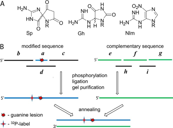
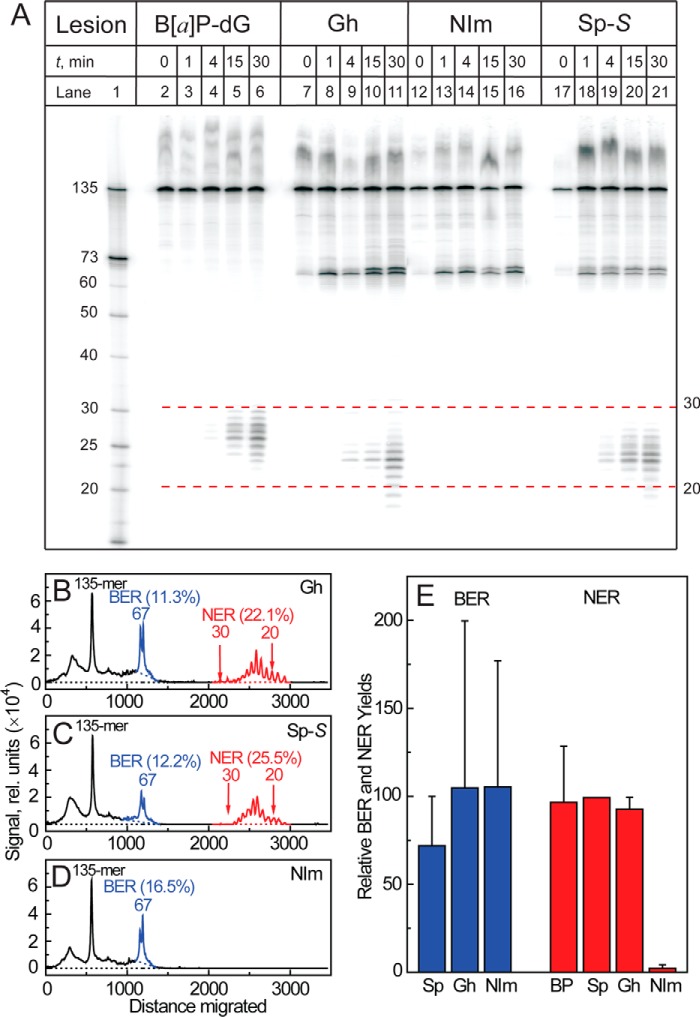
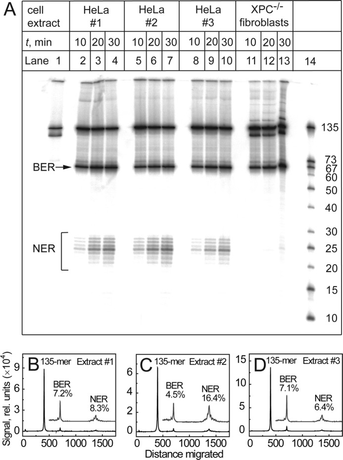
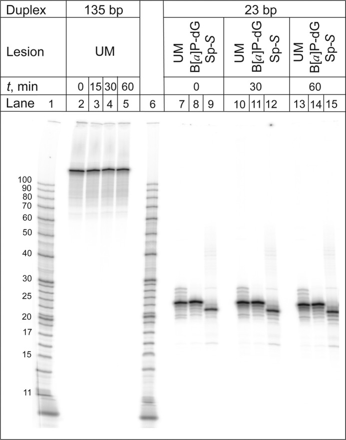
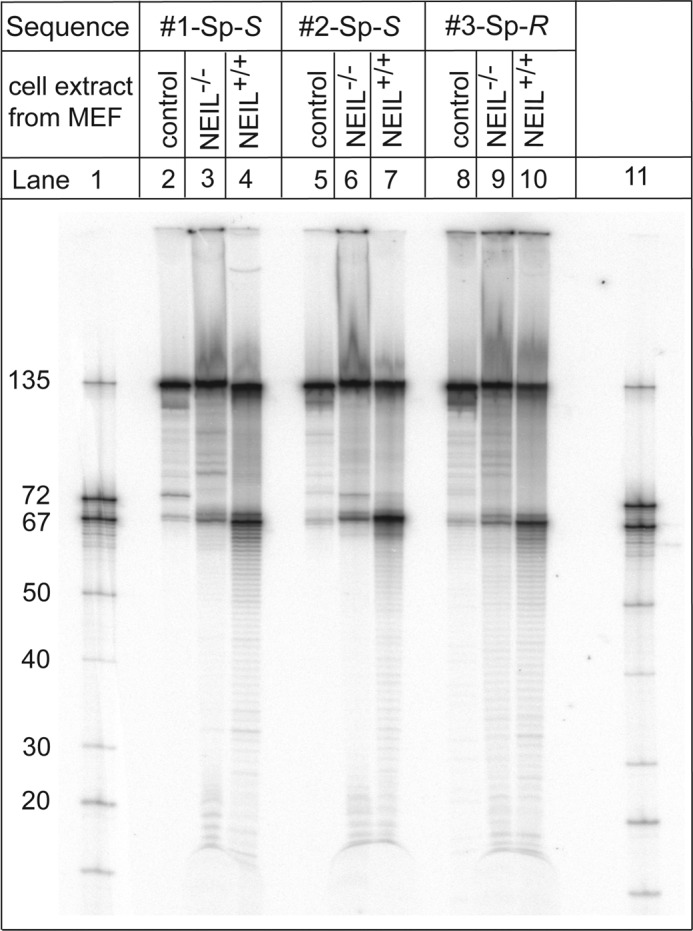
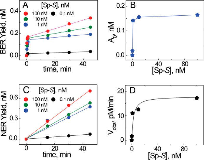
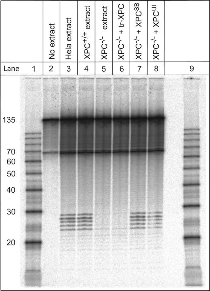
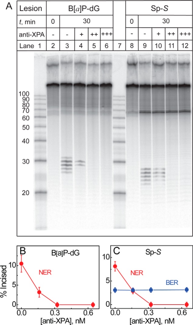
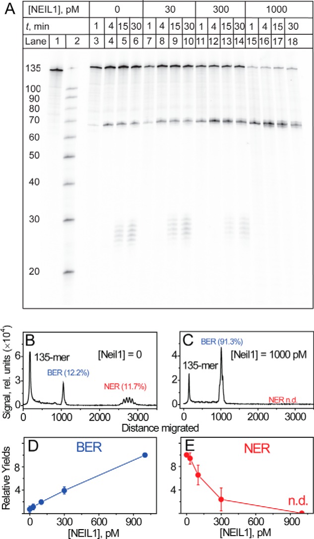
The well known biomarker of oxidative stress, 8-oxo-7,8-dihydroguanine, is more susceptible to further oxidation than the parent guanine base and can be oxidatively transformed to the genotoxic spiroiminodihydantoin (Sp) and 5-guanidinohydantoin (Gh) lesions. Incubation of 135-mer duplexes with single Sp or Gh lesions in human cell extracts yields a characteristic nucleotide excision repair (NER)-induced ladder of short dual incision oligonucleotide fragments in addition to base excision repair (BER) incision products. The ladders were not observed when NER was inhibited either by mouse monoclonal antibody (5F12) to human XPA or in XPC(-/-) fibroblast cell extracts. However, normal NER activity appeared when the XPC(-/-) cell extracts were complemented with XPC-RAD23B proteins. The Sp and Gh lesions are excellent substrates of both BER and NER. In contrast, 5-guanidino-4-nitroimidazole, a product of the oxidation of guanine in DNA by peroxynitrite, is an excellent substrate of BER only. In the case of mouse embryonic fibroblasts, BER of the Sp lesion is strongly reduced in NEIL1(-/-) relative to NEIL1(+/+) extracts. In summary, in human cell extracts, BER and NER activities co-exist and excise Gh and Sp DNA lesions, suggesting that the relative NER/BER product ratios may depend on competitive BER and NER protein binding to these lesions.

Keywords: DNA damage; DNA repair; base excision repair (BER); nucleotide excision repair; oxidative stress.


Figures

FIGURE 1.
FIGURE 1.
BER and NER substrates. A, structures of the oxidatively generated Sp, Gh, and NIm lesions. B, construction of the 32P-internally labeled 135-bp duplexes containing site-specifically positioned guanine lesions. a, 11-mer 5′-CCATCXCTACC, where X = Sp-S, Gh, NIm, cis-B[a]P-N2-dG. b, 62-mer 5′-GACCTGAACACGTACGGAATTCGATATCCTCGAGCCAGATCTGCGCCAGCTGGCCACCC CCA. c, 62-mer 5′-ACCCGCCAAGCTTGGGCTGCAGCAGGTCGACTCTAGAGGATCCCGGGCGAGCTCGAATTCGC. d, 31-mer 5′-CTTGGCGCTCGGTAGCGATGGTCAGGGTGGC. e, 56-mer 5′-GCGAATTCGAGCTCGCCCGGGATCCTCTAGAGTCGACCTGCTGCAGCCCAAGCTTG. f, 27-mer 5′-GCGGGTGGTAGCGATGGTGGGGGTGGC. g, 52-mer 5′-CAGCTGGCGCAGATCTGGCTCGAGGATATCGAATTCCGTACGTGTTCAGGTC. h, 20-mer 5′-TACCACCCGCCAAGCTTGGG. i, 20-mer 5′-GCGCCAGCTGGCCACCCCCA.
FIGURE 2.
FIGURE 2.
BER incisions and NER dual incisions of 135-mer DNA duplexes with Gh, Sp-S, NIm, or B[a]P-dG lesions in cell-free extracts from HeLa cells. A, representative denaturing gel showing the appearance of excision (BER) and dual incision (NER) products elicited by the Gh, Sp-S, and NIm-containing 135-bp duplexes (1 nm) as a function of incubation time in the same HeLa cell extract (0, 1, 4, 15, and 30 min). The 10R (+)-cis-anti-B[a]P-N2-dG adducts were used as positive controls of NER activity. Lane 1, oligonucleotide size markers. The apparent size range of the NER dual incision products is shown by dotted lines (red). B–D, histograms derived from the gel autoradiograph (A, lanes 11, 16, and 21) depicting the relative distributions of BER and NER excision products in the 135-bp duplexes containing Gh (B), Sp-S (C), and NIm (D) lesions after incubation in the same HeLa cell extracts for 30 min. E, average NER (red) and BER (blue) yields based on three independent experiments, each in a different cell extract measured after a 30-min incubation period. In each of these individual experiments, the NER yields of Gh, NIm, and B[a]P-dG and the BER yields of Sp-S, Gh, and NIm lesions were normalized to the Sp-S NER value (arbitrarily assigned the value of 100) in the same experiment. The blue and red bars represent the averages of the three independent experiments with their standard deviations. Other details are described in “Experimental Procedures.”
FIGURE 3.
FIGURE 3.
BER and NER of Sp-R lesions in HeLa cell extracts and XPC-proficient or XPC-deficient fibroblasts. A, three additional examples of single (BER) and dual (NER) incisions elicited by the stereoisomeric Sp-R lesions containing 135-bp duplexes as a function of incubation time (10, 20, and 30 min) in HeLa cell extracts (three different preparations) and extracts from XPC-deficient (XPC−/−) cells. Lane 14, oligonucleotide size markers. B–D, histograms derived from the gel autoradiograph (A) depicting the relative distributions of BER and NER excision products in the 135-bp Sp-R duplexes after incubation in three different preparations of HeLa cell extracts for 30 min.
FIGURE 4.
FIGURE 4.
Denaturing gel showing different electrophoretic mobilities of single-stranded 23-mer oligonucleotides without (unmodified) and with single B[a]P-dG adducts or Sp-S lesions. In total, eight such experiments were performed that showed qualitatively similar behavior. The 23-bp duplexes were constructed from 5′-TGGCCACCCTGA*CCATCXCTACC oligonucleotides with the 32P internal label at position indicated by an asterisk (*) and X = G, B[a]P-dG, or Sp-S. These 23-mer sequences (1 nm) were annealed with their fully complementary 23-mer strands containing a C opposite X in the modified strands, incubated in HeLa cell extracts with an excess of the unlabeled 135-bp duplexes (10 nm) for fixed periods of time (0, 30, and 60 min), and subsequently analyzed by denaturing gel electrophoresis. Lanes 1 and 6, oligonucleotide size markers. UM, unmodified 135-bp duplex control.
FIGURE 5.
FIGURE 5.
BER activities of 135-bp duplexes with centrally positioned Sp lesions in cell-free extracts from wild (NEIL1+/+) and KO (NEIL1−/−) MEFs. The 135-mer modified strands contained the centrally positioned sequences: 5-CCATC(Sp-S)CTACC (#1), 5′-CCACCAAC(Sp-S)CTACCACC (#2), or 5′-CCACCAAC(Sp-R)TCACCACC (#3); the remaining base sequences in the 135-mer duplexes with #2 and #3 are shown in Fig. 1B. Lanes 1 and 11, oligonucleotide size markers. The BER activities (yield of 67-mer fragments) are strongly reduced in NEIL1−/− sequences in all three sequence contexts.
FIGURE 6.
FIGURE 6.
Example of the kinetics of appearance of BER and NER incision product formation (A and C) and the effects of DNA substrate concentration (B and D). The DNA substrate was a 135-bp duplex containing a single Sp-S lesions in HeLa cell extracts. Similar qualitative behavior was observed with four other cell extracts analyzed in detail, although the BER product yields were variable as discussed in connection with the error bars shown in Fig. 2E and the results in Fig. 3, B–D. A, time course of single incision 67-mer BER product formation. The solid lines are the least-squares fits of the BER Equation 1 to the experimental data points. B, dependence of the burst amplitude on the DNA substrate concentration; the solid line is meant to guide the eye only. C, time course of dual incision NER product formation. The solid lines are the least-squares linear fits to the data points. D, dependence of the dual excision yield at the 30-min incubation time point on the DNA substrate concentration. The solid line is a fit of the Michaelis-Menten equation, Vobs = Vmax[DNAS]/(Km + [DNAS]) with Vmax = pm·min−1, and Km = 2.5 nm to the observed NER rates.
FIGURE 7.
FIGURE 7.
XPC-RAD23B complementation experiments in XPC-deficient fibroblasts. Representative denaturing gel electrophoresis of NER products after incubation of Sp-S-containing 135-bp duplexes (1 nm) in normal HeLa cell extracts (lane 3), extracts from XPC+/+ fibroblasts (lane 4), extracts from XPC−/− cells (lane 5), extracts from XPC−/− cells complemented with 4 nm concentrations of truncated XPC-RAD23B (tr-XPC, lane 6), full XPC-RAD23B from Stony Brook University (XPCSB, lane 7), or from the University of Illinois (XPCUI, lane 8). Lanes 1 and 9, oligonucleotide size markers. The incubation times were 30 min in all experiments. In total, four such experiments were performed that showed qualitatively similar behavior.
FIGURE 8.
FIGURE 8.
Inhibition of NER activities of HeLa cell extracts by the addition of mouse monoclonal anti-XPA antibodies. A, denaturing gel showing the inhibition of NER dual incision activity associated with 135-bp Sp-S-containing duplexes (1 nm) as a function of a mouse monoclonal anti-XPA (5F12) concentration after incubation in HeLa cell extracts for fixed periods of time (0 and 30 min). The 10R (+)-cis-anti-B[a]P-N2-dG adduct embedded in 135-mers was used as a positive control of NER activity. Lanes 1 and 7, oligonucleotide size markers. B and C, average yields of BER (blue) and NER (red) BER incision and NER excision products measured at the 30-min incubation time point as a function of anti-XPA concentration (averages of three independent experiments).
FIGURE 9.
FIGURE 9.
Competition between BER and NER pathways in HeLa cell extracts. A, autoradiograph of a denaturing gel showing the BER and NER product yields as a function of the concentration of the BER glycosylase NEIL1. The substrates were 135-bp duplexes (1 nm) containing single Sp-S lesions. Lane 1, DNA substrate not treated in cell extracts (control). Lane 2, oligonucleotide size markers. B and C, histograms derived from the gel autoradiograph (A, lanes 6 and 18 at the 30-min incubation time point). D, average NER (red) and BER (blue) yields based on three independent experiments, each in different cell extracts measured after a 30-min incubation period. In each of these individual experiments the NER and BER yields of Sp-S were normalized either to the NER value at [NEIL1] = 0 or the BER value at [NEIL1] = 1000 pm (arbitrarily assigned a value of 10 in each case).

References

    1. Lonkar P., and Dedon P. C. (2011) Reactive species and DNA damage in chronic inflammation: reconciling chemical mechanisms and biological fates. Int. J. Cancer 128, 1999–2009 - PMC - PubMed
    1. Coussens L. M., and Werb Z. (2002) Inflammation and cancer. Nature 420, 860–867 - PMC - PubMed
    1. Grivennikov S. I., Greten F. R., and Karin M. (2010) Immunity, inflammation, and cancer. Cell 140, 883–899 - PMC - PubMed
    1. Kidane D., Chae W. J., Czochor J., Eckert K. A., Glazer P. M., Bothwell A. L., and Sweasy J. B. (2014) Interplay between DNA repair and inflammation, and the link to cancer. Crit. Rev. Biochem. Mol. Biol. 49, 116–139 - PMC - PubMed
    1. Cadet J., Douki T., and Ravanat J. L. (2008) Oxidatively generated damage to the guanine moiety of DNA: mechanistic aspects and formation in cells. Acc. Chem. Res. 41, 1075–1083 - PubMed

Publication types

LinkOut - more resources