

Comment
doi: 10.1038/nsmb.3157.
The mystery of the fusion pore
Affiliations
- PMID: 26733219
- PMCID: PMC4910880
- DOI: 10.1038/nsmb.3157
Item in Clipboard
Comment
The mystery of the fusion pore
Nat Struct Mol Biol.
2016 Jan.
No abstract available
Figures
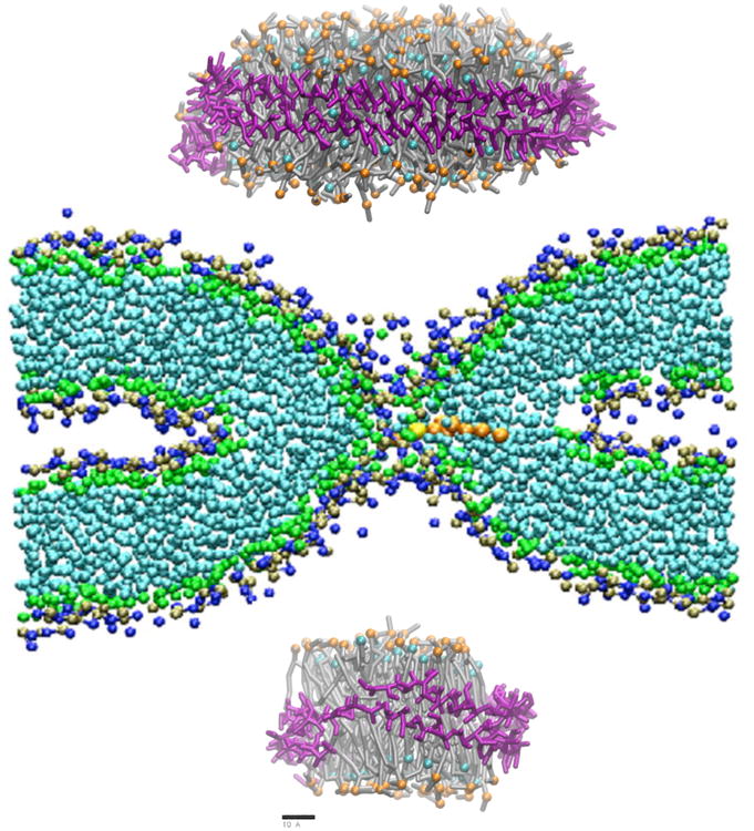
A lipidic fusion pore (center) might fit a 12 nm nanodiscs (top) but not a 6 nm nanodisc (bottom). Nanodiscs were simulated using GROMACS 4.6 using Martini force field . The structure of ∼12nm MSP1E2 was modeled based on crystal structure 1AV1 of the lipid binding domain of ApoA-I. To generate the smaller 6 nm disc helix 4 to helix 6 were deleted using Modeler. The lipids were chosen based on the synaptic vesicle lipid composition (12 nm disc: 154 CHOL, 13 PPCS, 69 POPC, 89 POPE and 25 POPS; 6 nm disc: 50 CHOL, 5 DPSM (sphingomyelin), 23 POPC, 30 POPE, 9 POPS). Lipid numbers were chosen based on simulation results showing that MSP1E2 nanodisc contains ∼125 DMPC lipids/leaflet . DMPC has an area per lipid (APL) of 0.61 nm2. Considering the average APL of 0.44 nm2 for the multicomponent planar bilayer, total number is ∼173 lipids/leaflet. The appropriate lipids from a pre-equilibrated asymmetric bilayer were placed in the empty nanodisc. A short equilibration (50 ns) was carried out with the head groups restrained along Z-direction (normal to bilayer) followed by an unrestrained 500 ns simulation.

(A) Possible arrangement of a 12 nm nanodisc docked to a membrane via 4 SNARE complexes after 20 ns simulation time. (B) Fusion pore snapshot after 1.665 μs simulation time with the isodensity surface of waters in the pore in side view. Colors: Syb2 – blue, Stx1 – red, SNAP-25 – green, MSP - yellow.
Comment on
-
Exocytotic fusion pores are composed of both lipids and proteins.Nat Struct Mol Biol. 2016 Jan;23(1):67-73. doi: 10.1038/nsmb.3141. Epub 2015 Dec 14. Nat Struct Mol Biol. 2016. PMID: 26656855 Free PMC article.
References
Publication types
MeSH terms
Substances
Grants and funding
LinkOut - more resources
Full Text Sources
Other Literature Sources
