Review
. 2016 Jun 20:625:11-5.
doi: 10.1016/j.neulet.2015.12.012. Epub 2015 Dec 28.

Role of HDACs in optic nerve damage-induced nuclear atrophy of retinal ganglion cells

Affiliations
Review

Role of HDACs in optic nerve damage-induced nuclear atrophy of retinal ganglion cells

Heather M Schmitt et al. Neurosci Lett. .

Abstract

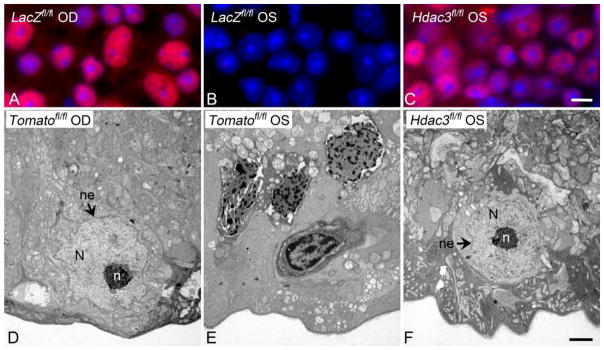
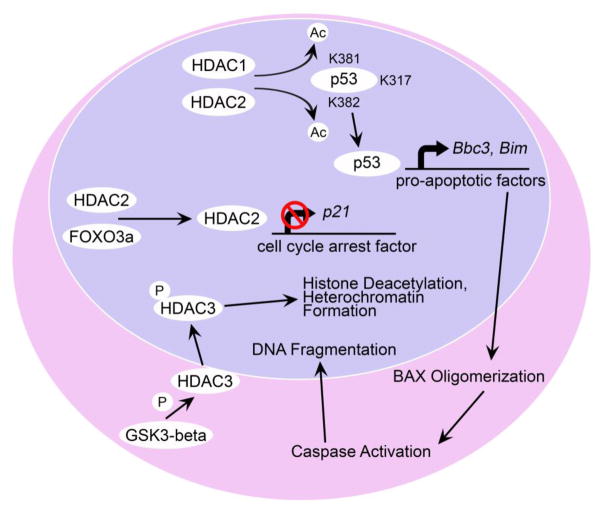
Optic neuropathies are characterized by retinal ganglion cell (RGC) death, resulting in the loss of vision. In glaucoma, the most common optic neuropathy, RGC death is initiated by axonal damage, and can be modeled by inducing acute axonal trauma through procedures such as optic nerve crush (ONC) or optic nerve axotomy. One of the early events of RGC death is nuclear atrophy, and is comprised of RGC-specific gene silencing, histone deacetylation, heterochromatin formation, and nuclear shrinkage. These early events appear to be principally regulated by epigenetic mechanisms involving histone deacetylation. Class I histone deacetylases HDACs 1, 2, and 3 are known to play important roles in the process of early nuclear atrophy in RGCs, and studies using both inhibitors and genetic ablation of Hdacs also reveal a critical role in the cell death process. Select inhibitors, such as those being developed for cancer therapy, may also provide a viable secondary treatment option for optic neuropathies.

Keywords: Apoptosis; Glaucoma; Heterochromatin; Histone deacetylase (HDAC); Neurodegeneration; Nuclear atrophy; Optic nerve injury; Retinal ganglion cell.


Figures

Figure 1
Figure 1. Conditional knockout of Hdac3 prevents heterochromatin formation after optic nerve crush
Conditional knockout of Hdac3 in RGCs was achieved by injecting AAV2-Cre virus into the left (OS) eye of Hdac3fl/fl mice prior to ONC. AAV2-Cre injected into the OS eyes of Rosa26-Tomatofl/fl mice served as virus-injected controls. Contralateral right (OD) eyes served as uncrushed and non-injected controls. Retinas were harvested at 5 days following crush injury for evaluation. (A) Retinal whole mount showing staining for acetylated histone H4 in unaffected OS eyes. (B) Crush elicits wide spread deacetylation of histones, but this process is blocked in RGCs lacking Hdac3 (C). DAPI counterstain. (Scale bar = 10 μm). (D) Transmission electron micrograph (TEM) image of a healthy cell in the ganglion cell layer (GCL) of a Rosa26-Tomatofl/fl control OD eye. (E) TEM image of heterochromatic cells in the GCL of an AAV2-Cre/GFP injected and crushed Rosa26-Tomatofl/fl OS eye. (F) TEM image of a cell in the GCL of the Hdac3 conditional knockout crushed OS eye. Healthy nuclei (N) are euchromatic and have well-formed nucleoli (n) and intact nuclear envelopes (ne), while injured cells exhibit heterochromatic nuclei with degrading nuclear envelopes. (Scale bar = 2μm)
Figure 2
Figure 2. Potential pathways of HDAC activity in neuronal toxicity
HDAC1 and HDAC2 reportedly activate p53 by deacetylating its K381 and K382 residues, where it leads to an increase in the expression of genes involved in apoptosis, including, Bbc3 (PUMA), and Bim [–30]. These BH3-only activator proteins then facilitate the activation of BAX, leading to caspase activity and the activation of an endonuclease that digests DNA. HDAC2 may have an additional function in silencing other p53-target genes, such as p21. In this mechanism, HDAC2 is associated with FOXO3a, which recruits it to the p21 promoter site, where it deacetylates histones in this region and represses transcription [34]. HDAC3 plays a primary role in histone deacetylation, and heterochromatin formation [3, 4]. In neurons, HDAC3 is activated by phosphorylation from GSK3β serine/threonine kinase, which becomes active when the PI3K/Akt signaling pathway is attenuated by loss of trophic factor support [26].

References

    1. McKinnon SJ, Schlamp CL, Nickells RW. Mouse models of retinal ganglion cell death and glaucoma. Exp Eye Res. 2009;88:816–824. - PMC - PubMed
    1. Janssen KT, Mac Nair CE, Dietz JA, Schlamp CL, Nickells RW. Nuclear atrophy of retinal ganglion cells precedes the bax-dependent stage of apoptosis. Invest Ophthalmol Vis Sci. 2013;54:1805–1815. - PMC - PubMed
    1. Pelzel HR, Schlamp CL, Nickells RW. Histone H4 deacetylation plays a critical role in early gene silencing during neuronal apoptosis. BMC Neurosci. 2010;11:62. - PMC - PubMed
    1. Schmitt HM, Pelzel HR, Schlamp CL, Nickells RW. Histone deacetylase 3 (HDAC3) plays an important role in retinal ganglion cell death after acute optic nerve injury. Mol Neurodegener. 2014;9:39. - PMC - PubMed
    1. Tiwari S, Dharmarajan S, Shivanna M, Otteson DC, Belecky-Adams TL. Histone deacetylase expression patterns in developing murine optic nerve. BMC Dev Biol. 2014;14:30. - PMC - PubMed

Publication types

Substances