. 2016 Jan 6:6:18668.
doi: 10.1038/srep18668.

RPS23RG1 reduces Aβ oligomer-induced synaptic and cognitive deficits

Affiliations

RPS23RG1 reduces Aβ oligomer-induced synaptic and cognitive deficits

Li Yan et al. Sci Rep. .

Abstract

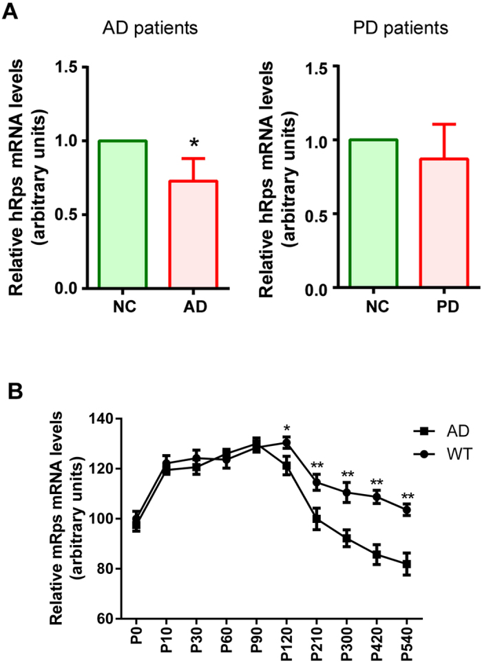
Alzheimer's disease (AD) is the most common form of dementia in the elderly. It is generally believed that β-amyloidogenesis, tau-hyperphosphorylation, and synaptic loss underlie cognitive decline in AD. Rps23rg1, a functional retroposed mouse gene, has been shown to reduce Alzheimer's β-amyloid (Aβ) production and tau phosphorylation. In this study, we have identified its human homolog, and demonstrated that RPS23RG1 regulates synaptic plasticity, thus counteracting Aβ oligomer (oAβ)-induced cognitive deficits in mice. The level of RPS23RG1 mRNA is significantly lower in the brains of AD compared to non-AD patients, suggesting its potential role in the pathogenesis of the disease. Similar to its mouse counterpart, human RPS23RG1 interacts with adenylate cyclase, activating PKA/CREB, and inhibiting GSK-3. Furthermore, we show that human RPS23RG1 promotes synaptic plasticity and offsets oAβ-induced synaptic loss in a PKA-dependent manner in cultured primary neurons. Overexpression of Rps23rg1 in transgenic mice consistently prevented oAβ-induced PKA inactivation, synaptic deficits, suppression of long-term potentiation, and cognitive impairment as compared to wild type littermates. Our study demonstrates that RPS23RG1 may reduce the occurrence of key elements of AD pathology and enhance synaptic functions to counteract oAβ-induced synaptic and cognitive deficits in AD.


Figures

Figure 1
Figure 1. Identification and cloning of the human RPS23RG1gene.
(A) Diagram showing human fetal brain cDNA library constructed in a phagemid vector. PCR primers used to amplify human RPS23RG1 gene are depicted by black arrows. (B) The full-length 1569bp human RPS23RG1 cDNA sequence was reconstituted from PCR products amplified from a human fetal brain phagemid cDNA library. The cDNA sequence was also confirmed by RT-PCR from total human fetal brain RNA. ATG start codon and TGA stop codon were shown in red. Intron and exon boundaries were shown in green. (C) The genomic contig of human RPS23RG1 in chromosome 8 and the encoded protein was shown. The transmembrane domain was indicated in blue.
Figure 2
Figure 2. PRS23RG1/Rps23rg1 mRNA levels are decreased in postmortem human AD patient and AD transgenic mouse brains.
(A) Quantification of RPS23RG1 (hRps) mRNA levels in the postmortem brains of human AD patients (left) or PD patients (right). Values were mean ± SEM (n = 8 for AD and n = 7 for PD, *p < 0.05 two-tailed Student’s t test). (B) The mRNA level of mouse Rps23rg1 (mRps) in Tg2576 AD or WT mouse brains at different ages were quantified for comparison (n = 4 at each data point, *p < 0.05, **p < 0.01, two-tailed Student’s t test).
Figure 3
Figure 3. Human RPS23RG1 overexpression reduces Aβ levels and tau-phosphorylation via its interaction with adenylate cyclase 8 and consequent PKA activation.
(A) Cells transfected with adenylate cyclase 8 (AC8) together with human RPS23RG1 (hRps) or mouse Rps23rg1 (mRps) were used for co-immunoprecipitation experiments. Cell lysates were incubated with mouse IgG (mIgG), anti-c-myc, rabbit IgG (rIgG), or anti-AC8. Immunoprecipitated proteins were subjected to western blot analysis using antibodies against AC8 or myc (for hRps or mRps, respectively). Co-IP experiments were reproduced 4 times. (B,C) Cells transfected with hRps, mRps, or control vector (Con) were analyzed for cAMP levels (B) or in vitro PKA activity (C). (D) Cells were transfected with hRps, mRps, or control vector (Con). Cell lysates were analyzed for phosphorylated and total GSK-3, phosphorylated and total CREB, CTF, and Rps (myc) levels. Conditioned media were analyzed using the 6E10 antibody to detect sAPPα levels. Summary graph showed relative p-GSK-3 and p-CREB levels. (E) Cells transfected with hRps (h), mRps (m), or control vector (V) were treated with DMSO (Con) or the PKA inhibitor H89. Cell lysates were analyzed for phosphorylated and total GSK-3 and Rps (myc) levels. Summary graph show relative p-GSK-3 levels. (F) Cells were transfected with hRps, mRps, or control vector (Con). Cell lysates were analyzed for phosphorylated (PHF-1 or p-tau) and total (tau) tau and Rps (myc). Summary graph showed relative p-tau levels normalized against total tau levels. (G) Conditioned media were also analyzed for Aβ42 secretion by ELISA as shown. All data were normalized to control values (as one arbitrary unit) and shown as mean ± SEM (n = 3, *p < 0.05, **p < 0.01, n.s.: not significant, one-way ANOVA with Dunnett’s multiple comparisons).
Figure 4
Figure 4. RPS23RG1overexpression mitigates oAβ-induced synaptic loss.
Cultured neurons were transfected with plasmids encoding human RPS23RG1 (hRps) and EGFP or EGFP alone, and then exposed to oAβs or control Aβ42-1. In some experiments, neurons were treated with additional DMSO or the PKA inhibitor H89. (A) Immunofluorescence images reveal localization of PSD-95 (red) and synapsin I (Syn; blue) clusters in neuronal dendrites. (B,C) Quantification of PSD-95 cluster (B) or PSD-95/Syn co-cluster (C) densities in neuronal dendrites (nEGFP/Ctrl = 12, nEGFP/Aβ = 10, nhRps/Ctrl = 11, nhRps/Aβ = 13). (D) Dendritic spines were visualized by transfected EGFP. (E) Quantification of spine density (nEGFP/Ctrl/DMSO = 11, nEGFP/Aβ/DMSO = 10, nhRps/Ctrl/DMSO = 11, nhRps/Aβ/DMSO = 13, nhRps/Ctrl/h89 = 10, nhRps/Aβ/H89 = 10). Scale bar, 5 μm. Values were mean ± SEM (n.s., not significant. *,#p < 0.05, **p < 0.01 one-way ANOVA with Dunnett’s multiple comparisons).
Figure 5
Figure 5. Rps23rg1knockdown aggravates oAβ-induced synaptic loss.
(A) Representative images showing expression of PSD-95 (green) in cultured neurons transfected with Rps23rg1 or control siRNA and exposed to oAβs or control Aβ42-1. Only neuronal dendrites transfected with Cy3-tagged siRNAs (not shown) were selected for analysis. (B) Quantification of PSD-95 cluster densities showed that Rps23rg1 knockdown aggravated oAβ-induced synaptic loss. (C) Dendritic spines were visualized by transfected EGFP. (D) Graph summary showing that oAβ-induced spine loss was further enhanced by Rps23rg1 siRNA. Scale bar, 5 μm. Values were mean ± SEM (n = 6 cells per group from 3 cultures). #p < 0.05, **,##p < 0.01 one-way ANOVA with Dunnett’s multiple comparisons.
Figure 6
Figure 6. Rps23rg1 verexpression alleviates oAβ-induced cognitive impairment in mice.
(A) Summary graph showing latency in finding hidden platforms during training sessions in Morris water maze tests. (B) Summary graph showing the percent of time spent in the target and the averaged percent of time spent in the other three quadrants during probe test. (C) Representative swimming patterns during probe test were shown. (D) Spontaneous alternations in Y-maze test. Rps23rg1 transgenic (Tg) mice showed improved performance compared to wild type (WT) mice in the presence and absence of oAβs. For both behavior tests, 11 WT/V, 10 WT/Aβ, 13 Tg/V, and 12 Tg/Aβ male mice at 2~3 months old were used, respectively. Values were mean ± SEM (n.s., not significant, *p < 0.05, **p < 0.01, ***p < 0.001 by two-way ANOVA with Dunnett’s multiple comparisons).
Figure 7
Figure 7. Rps23rg1 overexpression reduces oAβ-induced synaptic toxicity in mice.
(A) Golgi staining showing dendritic spines in the hippocampus of wild type (WT) or Rps23rg1 transgenic (Tg) mice injected with oAβs or vehicle. Scale bar, 5 μm. (B) Quantification of spine densities in (A). (C) Rps23rg1 overexpression prevented oAβ-induced PKA inactivation. PKA activity was determined in Rps23rg1 Tg and WT mice injected with oAβs or vehicle in the hippocampus. (D–L) Representative gel images (D) and summary bar graphs showing expression of p-GSK-3 (E), total GSK-3 (F), p-CREB (G), total CREB (H), PHF-1 p-tau (I), total tau (J), synaptophysin (SYP, K), and PSD-95 (L). Values were mean ± SEM (n = 4, n.s., not significant, *p < 0.05, **p < 0.01 one-way ANOVA with Dunnett’s multiple comparisons).
Figure 8
Figure 8. Rps23rg1overexpression alleviates oAβ-induced LTP suppression in the hippocampal CA1 region.
(A,B) Induction of LTP in CA1 was significantly attenuated in wild type (WT) mouse CA1, but not in Rps23rg1 transgenic (Tg) mouse CA1, following oAβ injection, when compared to WT mice injected with vehicle. In panel (B), horizontal line = 10 msec and vertical line = 0.2 mV. LTP was recorded on 4, 5, 7 slices from 3 WT/V, 4 WT/Aβ, and 6 Tg/Aβ mice, respectively. Values were mean ± SEM (*p < 0.05 by two-way ANOVA with Dunnett’s multiple comparisons).

Similar articles

Cited by

References

    1. Li Z. & Sheng M. Caspases in synaptic plasticity. Mol Brain 5, 15 (2012). - PMC - PubMed
    1. Tu S., Okamoto S. I., Lipton S. A. & Xu H. Oligomeric Aβ-induced synaptic dysfunction in Alzheimer’s disease. Mol Neurodegener 9, 48 (2014). - PMC - PubMed
    1. Terry R. D. et al. Physical basis of cognitive alterations in alzheimer’s disease: Synapse loss is the major correlate of cognitive impairment. Ann Neurol 30, 572–580 (1991). - PubMed
    1. Zemlan F. P. et al. Quantification of axonal damage in traumatic brain injury: affinity purification and characterization of cerebrospinal fluid tau proteins. J Neurochem 72, 741–50 (1999). - PubMed
    1. Kouri N. et al. Genome-wide association study of corticobasal degeneration identifies risk variants shared with progressive supranuclear palsy. Nat Commun 6, 7247 (2015). - PMC - PubMed

Publication types

MeSH terms