Case Reports
. 2016 Jan;14(1):8-17.
doi: 10.6004/jnccn.2016.0003.

A Distributed Network for Intensive Longitudinal Monitoring in Metastatic Triple-Negative Breast Cancer

Affiliations
Case Reports

A Distributed Network for Intensive Longitudinal Monitoring in Metastatic Triple-Negative Breast Cancer

C Anthony Blau et al. J Natl Compr Canc Netw. 2016 Jan.

Abstract

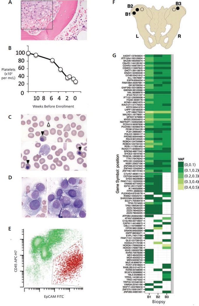
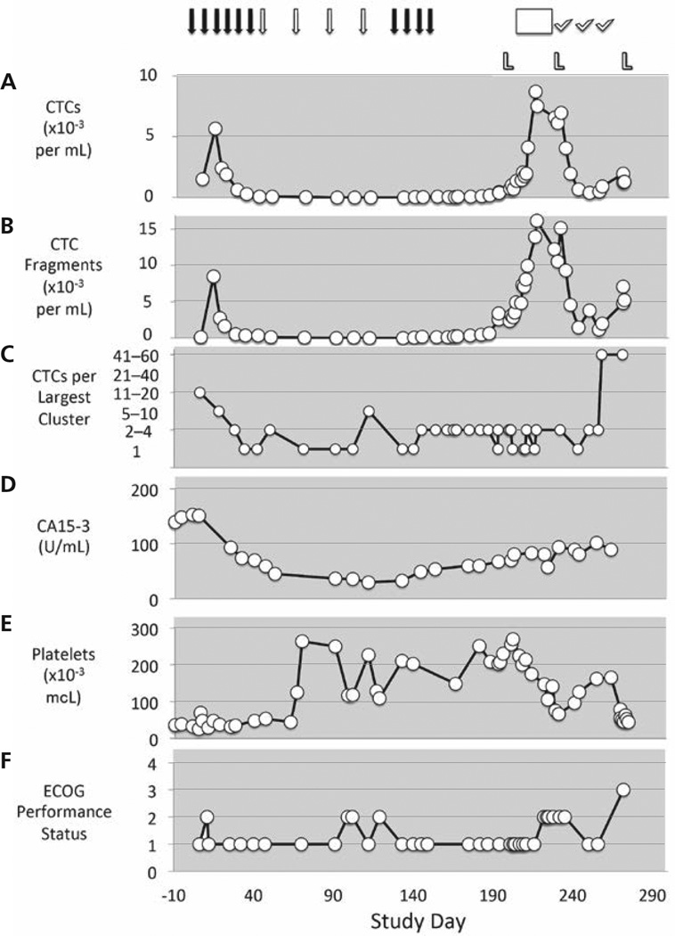
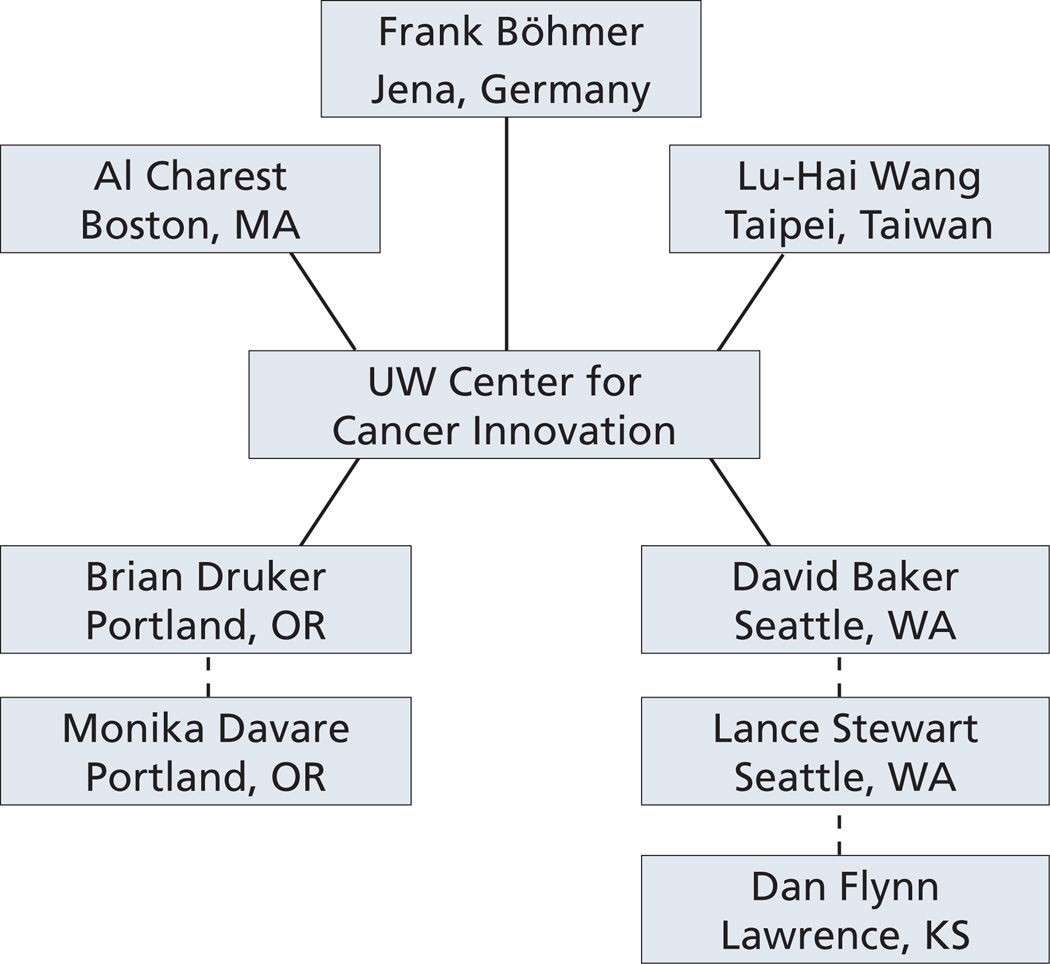
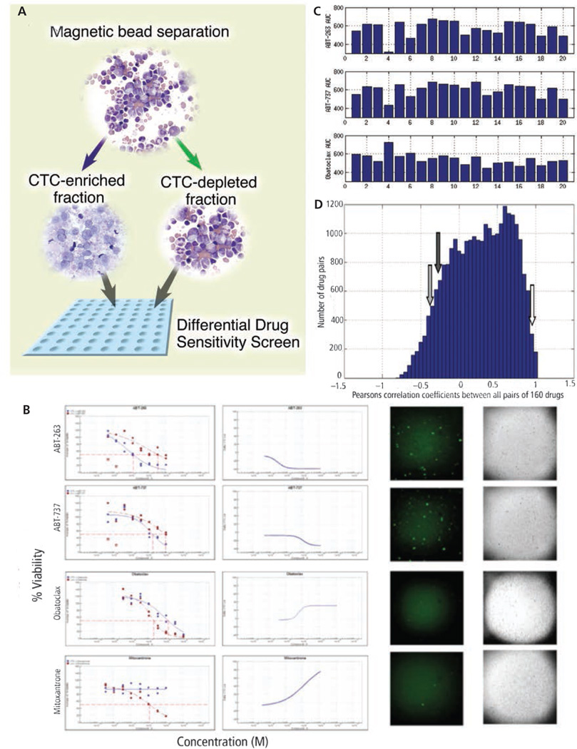
Accelerating cancer research is expected to require new types of clinical trials. This report describes the Intensive Trial of OMics in Cancer (ITOMIC) and a participant with triple-negative breast cancer metastatic to bone, who had markedly elevated circulating tumor cells (CTCs) that were monitored 48 times over 9 months. A total of 32 researchers from 14 institutions were engaged in the patient's evaluation; 20 researchers had no prior involvement in patient care and 18 were recruited specifically for this patient. Whole-exome sequencing of 3 bone marrow samples demonstrated a novel ROS1 variant that was estimated to be present in most or all tumor cells. After an initial response to cisplatin, a hypothesis of crizotinib sensitivity was disproven. Leukapheresis followed by partial CTC enrichment allowed for the development of a differential high-throughput drug screen and demonstrated sensitivity to investigational BH3-mimetic inhibitors of BCL-2 that could not be tested in the patient because requests to the pharmaceutical sponsors were denied. The number and size of CTC clusters correlated with clinical status and eventually death. Focusing the expertise of a distributed network of investigators on an intensively monitored patient with cancer can generate high-resolution views of the natural history of cancer and suggest new opportunities for therapy. Optimization requires access to investigational drugs.


Figures

Figure 1
Figure 1
Assembly of a distributed network. Components in place for all patients enrolled in ITOMIC-001 are shown in black. Components added specifically for the patient presented here are shown in red. Arrow indicates timeline of the study from day 0 to the time of the patient’s death on day 276. Abbreviation: CTC, circulating tumor cell.
Figure 2
Figure 2
Clinical status at time of enrollment, bone marrow biopsies, and mutation profiles. (A) Extensive tumor involvement in a prestudy bone marrow biopsy (box). (B) Progressive decline in platelet counts in the weeks before study enrollment. (C) Nucleated red blood cells (closed arrowheads) and tear drop cells (open arrowheads), consistent with a myelophthisic process. (D) Presence of tumor cells in touch preparations from bone marrow cores obtained at study entry. (E) Representative flow cytometry profile of hematopoietic cells (green) versus tumor cells (red) in a baseline bone marrow sample. (F) Schematic depiction of biopsy sites (circles) from the left (L) and right (R) posterior iliac crest. Filled circles (designated B1, B2, and B3) indicate samples that were analyzed. (G) Schematic depiction of the mutational profile of bone marrow samples B1–B3 as determined by MuTect. Select genes are noted. Colors denote variant allele frequencies (VAF).
Figure 3
Figure 3
Dynamic changes in circulating tumor cells (CTCs) in response to therapeutic interventions. (A) CTCs; (B) CTC fragments, and (C) CTC clusters over the study period in the context of CA15-3 (D), platelet counts (E), and ECOG performance status (F). Solid arrows denote weekly cisplatin treatment. Open arrows denote cisplatin treatment every 3 weeks. L refers to leukapheresis procedures. Crizotinib treatment is noted by an open rectangle. Eribulin treatments are denoted by check marks.
Figure 4
Figure 4
Experts contacted regarding the ROS1(Y2092C) variant observed in this patient. Solid lines denote primary contacts.
Figure 5
Figure 5
Development of a differential high throughput drug screen. (A) Following leukapheresis, circulating tumor cells (CTCs) were partially enriched using lineage depletion, and both the CTC-enriched and CTC-depleted cells were tested in a high-throughput screen of 160 investigational and approved oncology drugs. (B) Responses to BCL-2 inhibitors (ABT-263, ABT-737, obatoclax) and mitoxantrone (used as a positive control). Left columns show responses among CTC-enriched cells (blue lines) and CTC-depleted cells (red lines). Left middle column depicts summation of effects on CTC-enriched versus CTC-depleted cells. A declining slope indicates relatively greater potency against neoplastic cells, a rising slope indicates relatively greater potency against normal hematopoietic cells, whereas a flat line indicates similar potencies against neoplastic and hematopoietic cells. Right middle column depicts FITC-labeled, apoptotic cells detected using an antibody (M30), and the far right column depicts the corresponding bright field images. (C) Responses (indicated by area under the curve [AUC]) to ABT-263, ABT-737, and obatoclax across 20 triple-negative breast cancer (TNBC) cell lines (designated 1–20). (D) All correlations among all 160 drugs in the screen across the 20 TNBC cell lines. Open arrow shows that the correlation between ABT-263 and ABT-737 ranks in the top 3% approximately, whereas neither drug correlates with obatoclax (gray and black arrows).
Figure 6
Figure 6
Circulating tumor cell (CTC) cluster late in the study and extensive tumor occlusion of the pulmonary arteriolar vasculature. (A) A CTC cluster from study day 271. Left panel shows a merged image; middle panel shows cytokeratin staining; right panel shows EpCAM staining (original magnification ×40). The blue color indicates DAPI (4’,6-diamidino-2-phenylindole)-stained nuclei. (B) A low-power view of lung tissue in which multiple vessels contain clusters of dark-staining cells (arrowheads) (hematoxylin-eosin [H&E], original magnification ×4). (C) Image shows no identified involvement of pulmonary lymphatics (H&E, original magnification ×10). (D) Images shows a pulmonary infarct (H&E, original magnification ×2). (E) Magnified view of Figure 6B (H&E, original magnification ×10). (F) In many of these arteries, there is near occlusion of the lumen by breast carcinoma cells, and intimal expansion by smooth muscle cells, myofibroblasts, and extracellular material (H&E, original magnification ×40). (G) A Movat stain revealing that the vessels are arteries (containing both internal and external elastic laminae) (H&E, original magnification ×40).

References

    1. Scannell JW, Blanckley A, Boldon H, Warrington B. Diagnosing the decline in pharmaceutical R&D efficiency. Nat Rev Drug Discov. 2012;11:191–200. - PubMed
    1. Blau CA, Liakopoulou E. Can we deconstruct cancer, one patient at a time? Trends Genet. 2013;29:6–10. - PMC - PubMed
    1. Nowell PC. The clonal evolution of tumor cell populations. Science. 1976;194:23–28. - PubMed
    1. Dexter DL, Kowalski HM, Blazar BA, et al. Heterogeneity of tumor cells from a single mouse mammary tumor. Cancer Res. 1978;38:3174–3181. - PubMed
    1. Stephens PJ, McBride DJ, Lin M, et al. Complex landscapes of somatic rearrangement in human breast cancer genomes. Nature. 2009;462:1005–1010. - PMC - PubMed

Publication types