. 2016 Jan 19;113(3):E291-9.
doi: 10.1073/pnas.1518634113. Epub 2016 Jan 5.

Highly efficient delivery of functional cargoes by the synergistic effect of GAG binding motifs and cell-penetrating peptides

Affiliations

Highly efficient delivery of functional cargoes by the synergistic effect of GAG binding motifs and cell-penetrating peptides

James E Dixon et al. Proc Natl Acad Sci U S A. .

Erratum in

Abstract

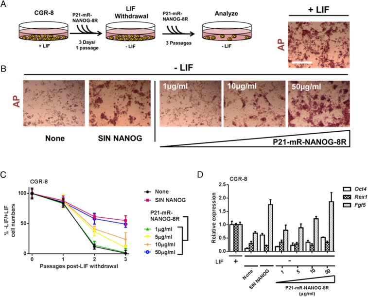
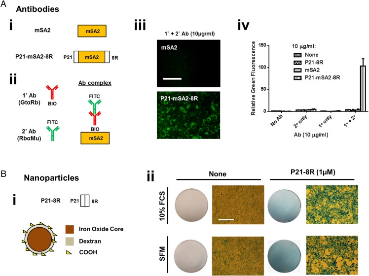
Protein transduction domains (PTDs) are powerful nongenetic tools that allow intracellular delivery of conjugated cargoes to modify cell behavior. Their use in biomedicine has been hampered by inefficient delivery to nuclear and cytoplasmic targets. Here we overcame this deficiency by developing a series of novel fusion proteins that couple a membrane-docking peptide to heparan sulfate glycosaminoglycans (GAGs) with a PTD. We showed that this GET (GAG-binding enhanced transduction) system could deliver enzymes (Cre, neomycin phosphotransferase), transcription factors (NANOG, MYOD), antibodies, native proteins (cytochrome C), magnetic nanoparticles (MNPs), and nucleic acids [plasmid (p)DNA, modified (mod)RNA, and small inhibitory RNA] at efficiencies of up to two orders of magnitude higher than previously reported in cell types considered hard to transduce, such as mouse embryonic stem cells (mESCs), human ESCs (hESCs), and induced pluripotent stem cells (hiPSCs). This technology represents an efficient strategy for controlling cell labeling and directing cell fate or behavior that has broad applicability for basic research, disease modeling, and clinical application.

Keywords: cell-penetrating peptides; differentiation; heparin-binding domain; human embryonic stem cells; transduction.


Conflict of interest statement

The authors declare no conflict of interest.

Figures

Fig. 1.
Fig. 1.
P21 improves PTD-mediated cellular uptake. (A) Schematic of the proteins created after screening domains that improve efficiency of protein delivery to cells. mR and mR-8R are described in Fig. S1. P21-mR is mRFP with an N-terminal fusion of the P21 domain of HB-EGF. P21-mR-8R is mRFP with N-terminal fusion of P21 and C-terminal fusion of 8R. (B) Fusion of P21 to mR-8R significantly improves uptake into NIH3t3 cells. Fluorescence microscopy images of NIH3t3 cells treated with proteins (20 µg/mL) for 12 h in standard media conditions. (Scale bar, 100 µm.) (C) P21-mR-8R is efficiently taken into hESCs and mESCs (HUES7 and CGR-8, respectively) and hiPSCs (IPS2) and mouse cardiomyocyte cell line HL1. Flow cytometry analyses of the mR-8R inefficiently delivered cell lines treated with proteins mR-8R (20 µg/mL) for 12 h. (D) P21-mR-8R initially strongly interacts with cell membranes and progressively is taken up and localized perinuclearly. Fluorescence (Top) and confocal laser scanning microscopy (Bottom) images of NIH3t3 cells treated with P21-mR-8R (20 µg/mL) for 1 h, 1 h with washes and a further 5 h incubation (in serum-free media), or 6 h treatment. Cells were preincubated for 1 h in serum-free media and transduced for the desired time in serum-free media. (Scale bars, top, 50 µm; bottom, 10 µm.) (E) Enhancement of cellular uptake mediated by P21 and 8R are affected by Trypsin proteolysis. Flow cytometry analyses NIH3t3 cells treated with proteins (20 µg/mL) for 1 h and a further 5 h incubation (in serum-free media), with or without 10 min predigestion with Trypsin or treatment with nonproteolytic cell dissociation solution (CDS). Cells were preincubated for 1 h in serum-free media, treated with Trypsin, and transduced for 1 h in serum-free media. (F) Cell surface interaction of P21-containing proteins is disrupted by Tritonx100 treatment. Flow cytometry analyses of NIH3t3 cells treated with proteins (20 µg/mL) for 1 h and a further 5 h incubation (in serum-free media) with 10 min pretreatment of PBS or PBS containing 0.1% (vol/vol) Tritonx100 (Tx100). Cells were preincubated for 1 h in serum-free media, treated with PBS or PBS with Tx100, and transduced for 1 h in serum-free media. Error bars indicate SD. n = 6.
Fig. 2.
Fig. 2.
GET of Cre recombinase. (A) Schematic of the construct created to mark Cre activity in cells. Cre-mediated excision of a transcriptional STOP region flanked by loxP sites induces the constitutive expression of eGFP. Pr, promoter; βGal, β-galactosidase; Neo, Neomycin phosphotransferase. The NIH3t3 LSL-eGFP cell line was created by transfection and selection of NIH3t3 cells. (B) eGFP expression in untreated NIH3t3 LSP-eGFP cells or those transduced with SIN Cre lentivirus. (Left) Fluorescence microscopy. (Right) Flow cytometry histogram of eGFP expression. (Scale bar, 50 µm.) (C) Scheme of testing transduction of Cre activity in NIH3t3 LSL-eGFP cells. Cells were transduced with Cre proteins for 1 h and washed and cultured for 2 d before analyses. (D and E) P21-mR-Cre-8R is efficiently transduced and recombines DNA. (D) Fluorescence microscopy images Cre-transduced NIH3t3 LSL-eGFP with the variety of dosages. (Scale bar, 50 µm.) (E) Flow cytometry analyses of NIH3t3 LSL-eGFP cells transduced for 1 h with mR-Cre, mR-Cre-8R, and P21-mR-Cre-8R at a variety of dosages (0, 1, 10, 100, and 500 µg/mL), washed and cultured for 2 d. Graph shows percentage recombination (i.e., percentage of eGFP positive from total cell population). Error bars indicate SD. n = 6.
Fig. 3.
Fig. 3.
GET of NANOG promotes the self-renewal of mouse embryonic stem cells. (A) Scheme of testing activity of transduced NANOG in CGR-8 cells. Cells were transduced with P21-mR-NANOG-8R proteins (0, 1, 10, and 50 µg/mL) for 3 consecutive days (1 passage, 1:3 split), passaged 1:3, and plated into growth media with P21-mR-NANOG-8R but lacking LIF (−LIF). Cells were fed daily with –LIF media containing P21-mR-NANOG-8R and passaged 1:3 every 3 d for 2 passages (a total of 3 passages –LIF). (B) P21-mR-NANOG-8R rescues self-renewal of mESCs lacking LIF dose-dependently. AP staining of CGR-8 cells treated with P21-mR-NANOG-8R proteins and LIF withdrawal. AP activity and colony morphology is retained in CGR-8 cells cultured in LIF or without LIF but supplemented with SIN NANOG (to overexpress NANOG) or transduced with P21-mR-NANOG-8R. (Scale bar, 100 µm.) (C) P21-mR-NANOG-8R maintains the proliferation of mESCs lacking LIF dose-dependently. Percentage of the number of CGR-8 cells cultured without LIF versus those with LIF (percentage −LIF/+LIF) at passaging. In LIF-deficient CGR-8 cultures, proliferation is promoted when supplemented with SIN NANOG (to overexpress NANOG) or transduced with P21-mR-NANOG-8R. Error bars indicate SD. (D) NANOG-dependent rescue in LIF-deficient cultures generates a more epiblast-like gene expression profile. Relative gene expression analyses of LIF-deficient CGR-8 cultures using quantitative PCR. Cultures supplemented with SIN NANOG (to overexpress NANOG) or transduced with P21-mR-NANOG-8R have increased Fgf5 expression, reduced Rex1 expression, and retain Oct4 expression. Error bars indicate SE. n = 6.
Fig. 4.
Fig. 4.
GET of MYOD promotes myogenic differentiation of hESCs. (A) Scheme of testing the differentiation activity of transduced MYOD in HUES7 cells. HUES7 cells were plated onto gelatinized plastic and cultured in DMEM containing 10% (vol/vol) FCS. Cells were fed daily with DMEM containing 10% (vol/vol) FCS and P21-mR-MYOD-8R (0, 1, 5, 10, or 50 µg/mL) for 7 d. Media was then changed to DMEM containing 2% (vol/vol) horse serum (HS), human recombinant insulin, and P21-mR-MYOD-8R and fed daily for 3 d. (B–F) P21-mR-MYOD-8R drives myogenic differentiation of HUES7 cells to multinucleated Myotubes. (B) Light microscopy of HUES7 cells cultured under the myogenic regime supplemented with SIN MYOD (to overexpress MYOD) or transduced with P21-mR-MYOD-8R. Elongated fused Myotubes and single myocytes are generated with SIN-MYOD or high doses of P21-mR-MYOD-8R. (Scale bar, 100 µm.) (C) MYOD-dependent myogenic differentiation of hESCs. Relative gene expression analyses of HUES7 cultures using quantitative PCR. Cultures supplemented with SIN MYOD (to overexpress MYOD) or transduced with P21-mR-MYOD-8R have increased endogenous MYOD expression and skeletal muscle-specific ACTA1 expression. Error bars indicate SE. (D and E) P21-mR-MYOD-8R differentiated cells are multinucleated. (D) Quantitation of mean nuclei number per cell using PI staining. Error bars indicate SD. (E) Fluorescence microscopy images of HUES7 cells differentiated with P21-mR-MYOD-8R (50 µg/mL) and stained with nuclear dye DAPI. (Scale bar, 50 µm.) (F) P21-mR-MYOD-8R differentiated cells are MYOGENIN-positive. Quantitation of the percentage of MYOGENIN-positive cells using immunolabeling. Error bars indicate SD. n = 6.
Fig. 5.
Fig. 5.
GET of antibodies and nanoparticles. (A) GET of biotinylated cargoes using monomeric streptavidin (mSA2). (A, i) Schematic of the mSA2 proteins engineered to bind to and enable the uptake of biotinylated cargoes. We used P21-8R as a noninteracting control, mSA2 as a nontransducing control, and P21-mSA2-8R as the test protein. (A, ii) Schematic of the antibody (Ab) complexes of a biotinylated primary (1°) antibody (goat anti-rabbit; GtαRb) bound to an FITC-conjugated secondary (2°) antibody (rabbit anti-mouse; Rb αMu) used to test activity. (A, iii) GET delivery of Ab complexes was visible by fluorescence microscopy. (Scale bar, 50 µm.) With coincubation of P21-mSA2-8R (Bottom, 10 µg/mL), Ab complexes were efficiency delivered to cells. (A, iv) Flow cytometry demonstrating that 1°/2° Ab complexes (1 µg/mL) are taken into NIH3t3 cells poorly by direct incubation or when coincubated with mSA2 only. (B) GET of magnetic nanoparticles. (B, i) Schematic of the P21-8R peptide synthesized and test MNPs. We tested 250-nm Nanomag-D dextran shell/iron oxide core MNPs and conjugated P21-8R peptide to surface COOH groups. (B, ii) MNPs are taken into NIH3t3 cells most efficiently in serum-free media (SFM; Left). Light microscopy images of Prussian blue iron-stained NIH3t3 cells treated with MNPs (50 µg/mL) for 12 h in standard media conditions [10% (wt/vol) FCS] or SFM. Conjugation of P21-8R to MNPs significantly increases cellular uptake in both 10% (wt/vol) FCS and SFM conditions (circular image is of entire well). (Scale bar, 100 µm.) n = 6.
Fig. 6.
Fig. 6.
GET of nucleic acids. (A) Schematic of the LK15 proteins engineered to bind to and transduce nucleic acids. (B) Transfection of human mesenchymal stem cells using GET-LK15. Initially we assessed binding capacity of LK15 peptides for plasmid (p)DNA (SIN GFP, to express GFP on transfection), modified synthetic messenger RNA (modRNA) (Miltenyi Biotech; to express GFP on transfection), and small inhibitory (si)RNAs (labeled with FAM fluorophore to detect delivery). After optimizing ratios, we transfected human mesenchymal stem cells with P21-LK15-8R and pDNA (10 µg), modRNA (10 µg), or siRNA (1 µg) and visualized transfection by fluorescence microscopy. (Scale bar, 100 µm.) (C) Quantification of GET-LK15 transfection of human mesenchymal stem cells by flow cytometry (percentage transfection efficiency or relative fluorescence for siRNA) compared with LIPO2000 as a commercial standard. Error bars indicate SD.

References

    1. Gump JM, Dowdy SF. TAT transduction: The molecular mechanism and therapeutic prospects. Trends Mol Med. 2007;13(10):443–448. - PubMed
    1. El-Andaloussi S, Holm T, Langel U. Cell-penetrating peptides: Mechanisms and applications. Curr Pharm Des. 2005;11(28):3597–3611. - PubMed
    1. Meade BR, Dowdy SF. Exogenous siRNA delivery using peptide transduction domains/cell penetrating peptides. Adv Drug Deliv Rev. 2007;59(2-3):134–140. - PubMed
    1. Thompson DB, Cronican JJ, Liu DR. Engineering and identifying supercharged proteins for macromolecule delivery into mammalian cells. Methods Enzymol. 2012;503:293–319. - PMC - PubMed
    1. Cronican JJ, et al. A class of human proteins that deliver functional proteins into mammalian cells in vitro and in vivo. Chem Biol. 2011;18(7):833–838. - PMC - PubMed

Publication types

MeSH terms