. 2015 Dec 16:6:1369.
doi: 10.3389/fmicb.2015.01369. eCollection 2015.

Helicobacter pylori ATCC 43629/NCTC 11639 Outer Membrane Vesicles (OMVs) from Biofilm and Planktonic Phase Associated with Extracellular DNA (eDNA)

Affiliations

Helicobacter pylori ATCC 43629/NCTC 11639 Outer Membrane Vesicles (OMVs) from Biofilm and Planktonic Phase Associated with Extracellular DNA (eDNA)

Rossella Grande et al. Front Microbiol. .

Abstract

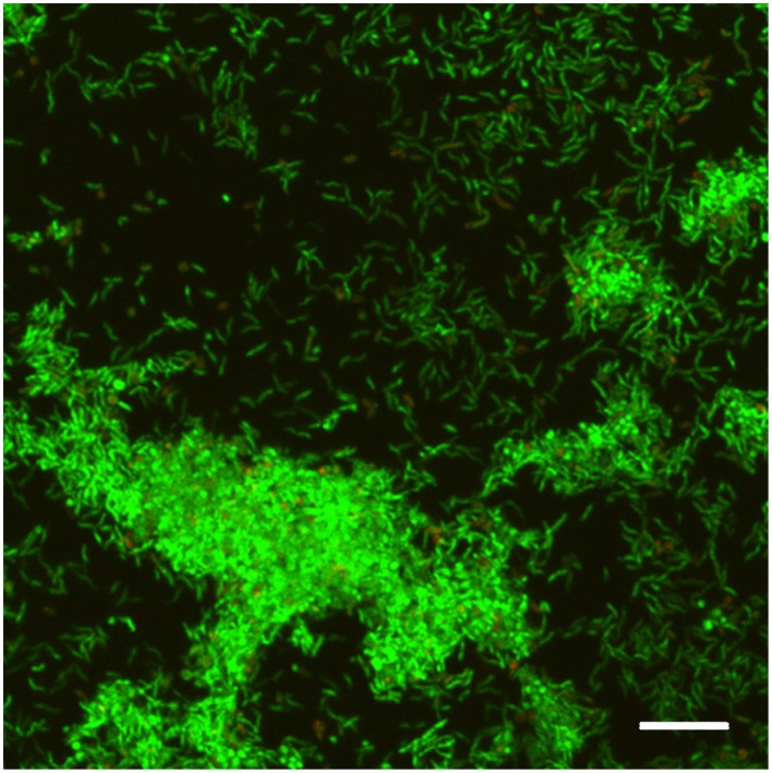
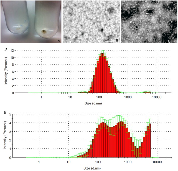
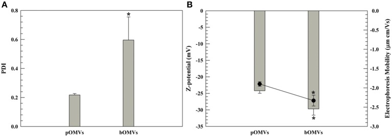
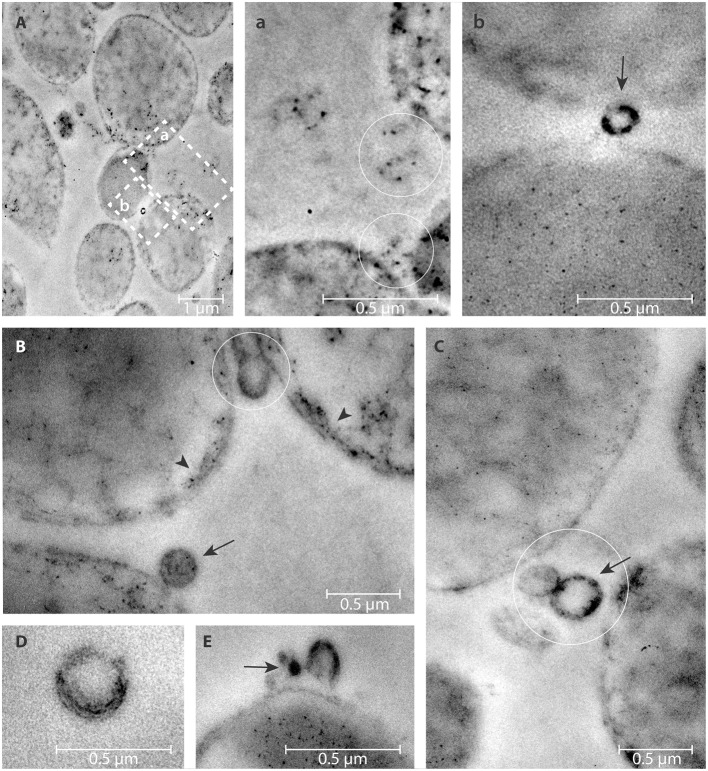
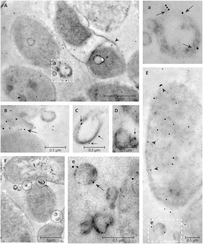
Helicobacter pylori persistence is associated with its capacity to develop biofilms as a response to changing environmental conditions and stress. Extracellular DNA (eDNA) is a component of H. pylori biofilm matrix but the lack of DNase I activity supports the hypothesis that eDNA might be protected by other extracellular polymeric substances (EPS) and/or Outer Membrane Vesicles (OMVs), which bleb from the bacteria surface during growth. The aim of the present study was to both identify the eDNA presence on OMVs segregated from H. pylori ATCC 43629/NCTC 11639 biofilm (bOMVs) and its planktonic phase (pOMVs) and to characterize the physical-chemical properties of the OMVs. The presence of eDNA in bOMVs and pOMVs was initially carried out using DNase I-gold complex labeling and Transmission Electron Microscope analysis (TEM). bOMVs and pOMVs were further isolated and physical-chemical characterization carried out using dynamic light scattering (DLS) analysis. eDNA associated with OMVs was detected and quantified using a PicoGreen spectrophotometer assay, while its extraction was performed with a DNA Kit. TEM images showed that eDNA was mainly associated with the OMV membrane surfaces; while PicoGreen staining showed a four-fold increase of dsDNA in bOMVs compared with pOMVs. The eDNA extracted from OMVs was visualized using gel electrophoresis. DLS analysis indicated that both planktonic and biofilm H. pylori phenotypes generated vesicles, with a broad distribution of sizes on the nanometer scale. The DLS aggregation assay suggested that eDNA may play a role in the aggregation of OMVs, in the biofilm phenotype. Moreover, the eDNA associated with vesicle membrane may impede DNase I activity on H. pylori biofilms. These results suggest that OMVs derived from the H. pylori biofilm phenotype may play a structural role by preventing eDNA degradation by nucleases, providing a bridging function between eDNA strands on OMV surfaces and promoting aggregation.

Keywords: Helicobacter pylori; biofilm; eDNA; nanoparticles; outer membrane vesicles (OMVs).


Figures

Figure 1
Figure 1
CLSM representative image of biofilm obtained from H. pylori ATCC 43629/NCTC 11639 samples stained with SYTO 9 (viable cells, green fluorescence) and Propidium Iodide (dead cells, red fluorescence) after 48 h of incubation. Scale bar = 5 μm. A representative image of five different experiments.
Figure 2
Figure 2
H. pylori ATCC 43629/NCTC 11639 pOMVs and bOMVs isolation. (A) OMVs ultra-centrifuged pellets from H. pylori planktonic and biofilm phases. The bOMVs pellets (left) were smaller than the pOMVs (right) and were white in color while the pOMVs pellets were dark yellow. (B,C) Negative staining of vesicles generated by H. pylori. DLS histogram analysis of pOMVs and bOMVs are shown in panels (D,E) respectively. (E) bOMVs had a more heterogeneous bimodal distribution of nanovesicles. In fact, there are three peaks, the first one at 681.1 nm (S.D. ± 256.2), the second one at 1760 nm (S.D. ± 2216) and the third one at 2557 nm. Figures are representative of five measurements.
Figure 3
Figure 3
DLS panel (PDI and Z-potential). PDI (A) and Z-potential (B) of pOMVs and bOMVs. (B), the histograms represent the Z-potential values (mV); while the black full circles are the electrophoretic mobility [(μm × cm)/Vs]. The sample analysis was carried out at 25°C and data are the average of 10 measurements ± standard deviation as triplicates. The error bar if not visible is within the symbol. *p ≤ 0.05.
Figure 4
Figure 4
DNase I-gold complexes labeling of 2-days old H. pylori ATCC 43629/NCTC 11639 mature biofilms. The colloidal gold labeled DNase I particles are used to detect the eDNA. The arrow represents the presence of eDNA on bOMVs membrane surface (b,B,C,E); while the arrow head represents the presence of eDNA on bacteria cell walls (B). OMVs are present between adjacent bacterial cells (circles, a,B,C). Representative arrangements between cells and vesicles are shown at higher resolution by the dashed squares “a” and “b” in (A). Panel (D) shows a single vesicle labeled with colloidal gold labeled DNase I particles.
Figure 5
Figure 5
DNase I-gold complexes labeling of H. pylori ATCC 43629/NCTC 11639 planktonic phase. The colloidal gold labeled DNase I particles are used to detect the eDNA. The arrow represents the presence of eDNA on pOMVs membrane surface (a,B,C,D,e); while the arrow head (A,E) represents the presence of eDNA on bacteria cell walls. OMVs appear between two cells (F, circles). The dashed squares (a,e), higher magnification of (A,E), highlight eDNA on the pOMVs surface.
Figure 6
Figure 6
eDNA associated with OMVs. (A) Detection and quantification of eDNA-pOMVs and eDNA-bOMVs associated, DNase I treated and untreated, by using PicoGreen assay. **p ≤ 0.003; *p ≤ 0.010 for differences of bOMVs compared to pOMVs. The protein content was used to normalize eDNA. (B) Representative image of agarose gel analysis of bOMVs-DNA from H. pylori ATCC 43629/NCTC 11639, untreated (lane 1), and DNase I-treated (lane 2). The lambda DNA/HindIII marker (M), DNA fragments are in base pairs.

References

    1. Allesen-Holm M., Barken K. B., Yang L., Klausen M., Webb J. S., Kjelleberg S., et al. . (2006). A characterization of DNA release in Pseudomonas aeruginosa cultures and biofilms. Mol. Microbiol. 59, 1114–1128. 10.1111/j.1365-2958.2005.05008.x - DOI - PubMed
    1. Barnes A. M., Ballering K. S., Leibman R. S., Wells C. L., Dunny G. M. (2012). Enterococcus faecalis produces abundant extracellular structures containing DNA in the absence of cell lysis during early biofilm formation. MBio 3:e00193–e00112. 10.1128/mbio.00193-12 - DOI - PMC - PubMed
    1. Bendayan M. (1981). Ultrastructural localization of nucleic acids by the use of enzyme-gold complexes. J. Histochem. Cytochem. 29, 531–541. 10.1177/29.4.6265546 - DOI - PubMed
    1. Carron M. A., Tran V. R., Sugawa C., Coticchia J. M. (2006). Identification of Helicobacter pylori biofilms in human gastric mucosa. J. Gastrointest. Surg. 10, 712–717. 10.1016/j.gassur.2005.10.019 - DOI - PubMed
    1. Celia C., Trapasso E., Locatelli M., Navarra M., Ventura C. A., Wolfram J., et al. . (2013). Anticancer activity of liposomal bergamot essential oil (BEO) on human neuroblastoma cells. Colloids Surf. B Biointerfaces 112, 548–553. 10.1016/j.colsurfb.2013.09.017 - DOI - PubMed

LinkOut - more resources