Recurrent arteriovenous malformation on palate after embolization combined surgical resection: preoperative magnetic resonance features and intraoperative angiographic findings
- PMID: 26734564
- PMCID: PMC4699938
- DOI: 10.5125/jkaoms.2015.41.6.346
Recurrent arteriovenous malformation on palate after embolization combined surgical resection: preoperative magnetic resonance features and intraoperative angiographic findings
Abstract
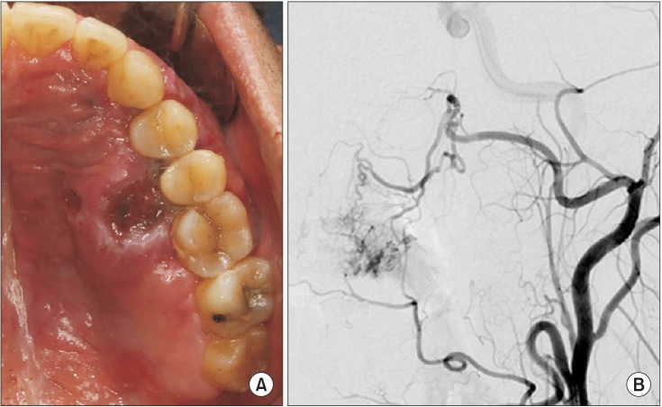
Angiography is the gold standard for the diagnosis and complete resection of arteriovenous malformations (AVMs). The absence of residual AVM after surgery is commonly believed to reduce the risk of future hemorrhage. However, AVMs can recur after proven complete angiographic resection can occur, albeit rarely, especially in the pediatric population. We report a rare case of a recurrent AVM two years after complete resection in an adult patient. This case report shows that AVMs in adults can recur despite their rarity and despite postoperative angiography confirming complete removal. Moreover, in this case, the recurrent AVM involved a new feeding vessel that was not involved with the initial lesion.
Keywords: Angiography; Arteriovenous malformations; Palate; Recurrence; Resection.
Conflict of interest statement
Figures
Similar articles
-
Intraoperative angiography of brain arteriovenous malformations.Neurosurgery. 1999 Sep;45(3):491-7; discussion 497-9. doi: 10.1097/00006123-199909000-00016. Neurosurgery. 1999. PMID: 10493371
-
Reappearance of arteriovenous malformations after complete resection of ruptured arteriovenous malformations: true recurrence or false-negative early postoperative imaging result?J Neurosurg. 2017 Apr;126(4):1088-1093. doi: 10.3171/2016.3.JNS152846. Epub 2016 May 27. J Neurosurg. 2017. PMID: 27231973
-
Recurrence of Cerebral Arteriovenous Malformations Following Resection in Adults: Does Preoperative Embolization Increase the Risk?Neurosurgery. 2016 Apr;78(4):562-71. doi: 10.1227/NEU.0000000000001191. Neurosurgery. 2016. PMID: 26702837
-
Spontaneous thrombosis of a residual arteriovenous malformation in eloquent cortex after surgery: case report.Neurosurgery. 2002 May;50(5):1142-5; discussion 1145-6. doi: 10.1097/00006123-200205000-00038. Neurosurgery. 2002. PMID: 11950420 Review.
-
How to deal with incompletely treated AVMs: experience of 67 cases and review of the literature.Acta Neurochir Suppl. 2011;112:123-9. doi: 10.1007/978-3-7091-0661-7_22. Acta Neurochir Suppl. 2011. PMID: 21692000 Review.
Cited by
-
Nasal septum angiofibroma: a rare condition with an unusual onset.J Korean Assoc Oral Maxillofac Surg. 2019 Feb;45(1):43-47. doi: 10.5125/jkaoms.2019.45.1.43. Epub 2019 Feb 26. J Korean Assoc Oral Maxillofac Surg. 2019. PMID: 30847296 Free PMC article.
-
Esthetic Root Coverage by Subepithelial Connective Tissue Graft with Management of Repeated Rupture of Palatal Arterial Bleeding: A Rare Case Report.J Pharm Bioallied Sci. 2021 Jun;13(Suppl 1):S861-S864. doi: 10.4103/jpbs.JPBS_550_20. Epub 2021 Jun 5. J Pharm Bioallied Sci. 2021. PMID: 34447216 Free PMC article.
-
Unique Case of Parathyroid Adenoma With Arteriovenous Malformation.Cureus. 2023 Jun 30;15(6):e41206. doi: 10.7759/cureus.41206. eCollection 2023 Jun. Cureus. 2023. PMID: 37525819 Free PMC article.
References
-
- Chiu YW, Wu HT, Chen YW, Lui MT, Kao SY, Lo WL. A giant venous malformation of face and neck: a case report. Taiwan J Oral Maxillofac Surg. 2011;22:110–117.
-
- Duncan IC, Fourie PA. Vascular malformations part 2: current classification of vascular malformations. South Afr J Radiol. 2004;8:23–30.
-
- Noreau G, Landry PP, Morais D. Arteriovenous malformation of the mandible: review of literature and case history. J Can Dent Assoc. 2001;67:646–651. - PubMed
-
- Sonstein WJ, Kader A, Michelsen WJ, Llena JF, Hirano A, Casper D. Expression of vascular endothelial growth factor in pediatric and adult cerebral arteriovenous malformations: an immunocytochemical study. J Neurosurg. 1996;85:838–845. - PubMed
Publication types
LinkOut - more resources
Full Text Sources
Other Literature Sources