. 2016 Jan 6;11(1):e0141028.
doi: 10.1371/journal.pone.0141028. eCollection 2016.

A Biomechanical Model of the Scapulothoracic Joint to Accurately Capture Scapular Kinematics during Shoulder Movements

Affiliations

A Biomechanical Model of the Scapulothoracic Joint to Accurately Capture Scapular Kinematics during Shoulder Movements

Ajay Seth et al. PLoS One. .

Abstract

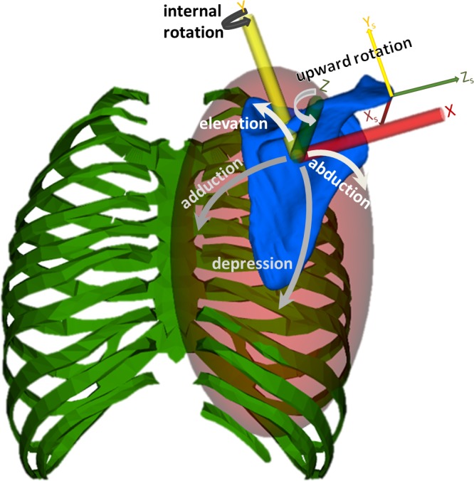
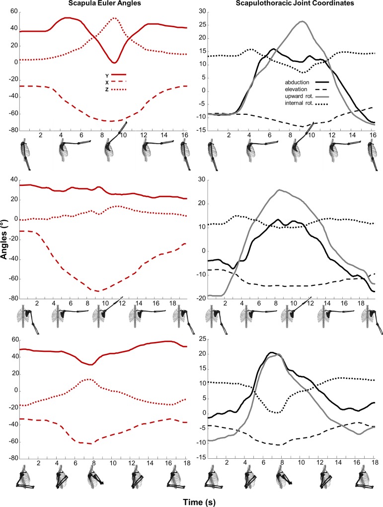
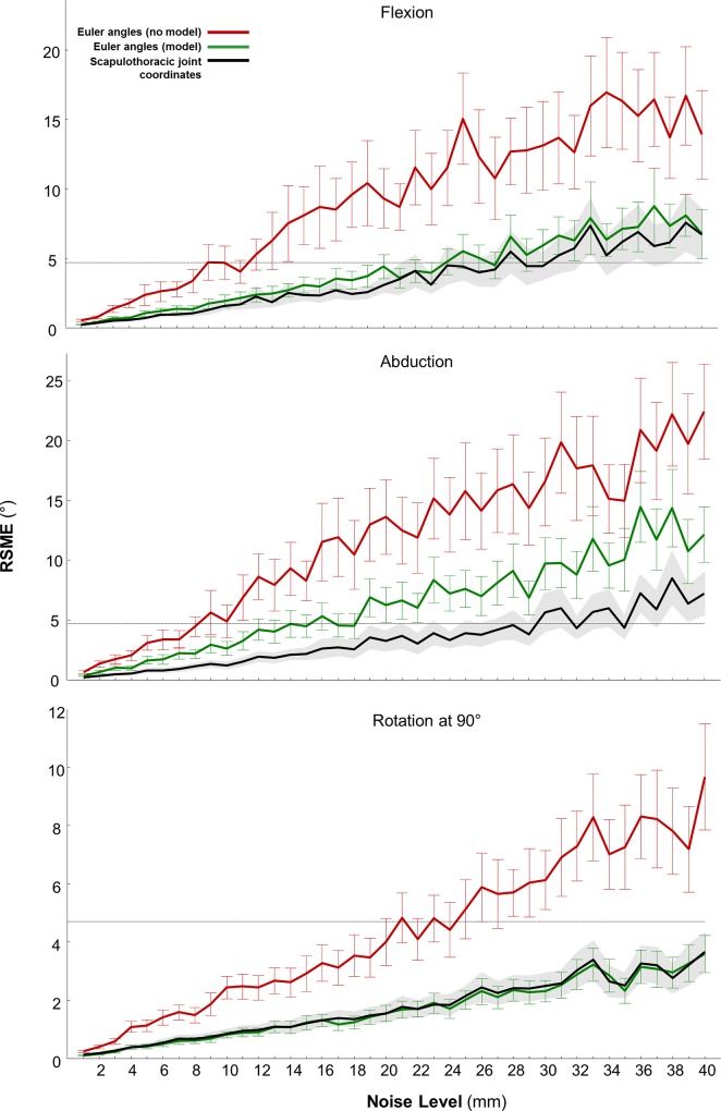
The complexity of shoulder mechanics combined with the movement of skin relative to the scapula makes it difficult to measure shoulder kinematics with sufficient accuracy to distinguish between symptomatic and asymptomatic individuals. Multibody skeletal models can improve motion capture accuracy by reducing the space of possible joint movements, and models are used widely to improve measurement of lower limb kinematics. In this study, we developed a rigid-body model of a scapulothoracic joint to describe the kinematics of the scapula relative to the thorax. This model describes scapular kinematics with four degrees of freedom: 1) elevation and 2) abduction of the scapula on an ellipsoidal thoracic surface, 3) upward rotation of the scapula normal to the thoracic surface, and 4) internal rotation of the scapula to lift the medial border of the scapula off the surface of the thorax. The surface dimensions and joint axes can be customized to match an individual's anthropometry. We compared the model to "gold standard" bone-pin kinematics collected during three shoulder tasks and found modeled scapular kinematics to be accurate to within 2 mm root-mean-squared error for individual bone-pin markers across all markers and movement tasks. As an additional test, we added random and systematic noise to the bone-pin marker data and found that the model reduced kinematic variability due to noise by 65% compared to Euler angles computed without the model. Our scapulothoracic joint model can be used for inverse and forward dynamics analyses and to compute joint reaction loads. The computational performance of the scapulothoracic joint model is well suited for real-time applications; it is freely available for use with OpenSim 3.2, and is customizable and usable with other OpenSim models.


Conflict of interest statement

Competing Interests: The authors have declared that no competing interests exist.

Figures

Fig 1
Fig 1. The four modeled degrees of freedom of the scapulothoracic joint.
The joint reference frame on the scapula (axes X,Y,Z) is used to locate the scapula with respect to the thorax. The joint reference frame on the scapula is computed according to the ISB recommendations [18] (shown as XS, YS, ZS), however our joint origin is located at the centroid of the anatomical markers used to define the joint frame instead of the Angulus Acromialis and its axes are rotated -90° about Y (to enable positive upward rotation about Z). The joint frame on the thorax defines the center of the scapulothoracic surface modeled as an ellipsoid (red shaded surface). Abduction (adduction) followed by elevation (depression) locate the joint frame origin of the scapula (blue) on the ellipsoid fixed to the parent thorax body (green). The scapula rotates upward (downward) about the normal to the surface (scapula Z-axis). Internal rotation or “winging” is a positive rotation about the Y-axis of the joint frame in the scapular plane, which remains tangent to the thoracic surface.
Fig 2
Fig 2. Scapula and Scapulothoracic joint kinematics during shoulder flexion, abduction, and rotation tasks.
Scapular kinematics described by the relative rotation of the scapula with respect to the thorax expressed as a body-fixed Y-X-Z Euler angle sequence according to the ISB standard (left panel: Y-internal rotation, solid; X-downward rotation, dashed; Z-posterior-tilting, dotted) and the scapulothoracic joint coordinates (right panel) with abduction (black solid), elevation (dashed), upward rotation (gray solid), and internal rotation or winging (dotted) reconstructed motion from measured bone-pin marker locations during shoulder tasks of: flexion, abduction, and rotation at 90° of humerus abduction.
Fig 3
Fig 3. Mean and standard deviation of root-mean-squared errors (RMSE) of scapular kinematics in the presence of noise compared to noise-free kinematics.
Scapular kinematics were computed with and without the scapulothoracic joint model during shoulder (humeral) flexion, abduction and rotation tasks. At every noise level, the model (green) and, in particular, the use of the scapulothoracic joint model coordinates (black), reduces RMSE by over 65% compared to direct scapula Euler angle calculations from markers (red). Standard deviations of Euler angles and joint coordinates are indicated by vertical bars and gray shading, respectively. The horizontal dotted line at 4.7° indicates where errors in scapular angles would result in an inability to distinguish the movement between different subjects (Bourne et al. 2011).
Fig 4
Fig 4. Scapulothoracic generalized coordinate forces (Nm) during shoulder flexion, abduction and rotation at 90° of shoulder abduction tasks.
Scapula abduction (bold), elevation (dashed), upward rotation (gray), and internal rotation (dotted) generalized torques computed from an inverse dynamics analysis. A large sustained torque is required to keep the scapula elevated against gravity and requires additional upward rotation torque (gray) to rotate the scapula and lift the humerus during arm elevation tasks.

References

    1. Veeger HEJ, van der Helm FCT (2007) Shoulder function: the perfect compromise between mobility and stability. J Biomech 40:2119–29 - PubMed
    1. Struyf F, Nijs J, Baeyens J-P, Mottram S, Meeusen R (2011) Scapular positioning and movement in unimpaired shoulders, shoulder impingement syndrome, and glenohumeral instability. Scand J Med Sci Sports 21:352–8 10.1111/j.1600-0838.2010.01274.x - DOI - PubMed
    1. Srikumaran U, Wells J, Freehill M (2014) Scapular Winging: A Great Masquerader of Shoulder Disorders. J Bone Jt Surg 122:1–13 - PubMed
    1. Lawrence RL, Braman JP, Laprade RF, Ludewig PM (2014) Comparison of 3-dimensional shoulder complex kinematics in individuals with and without shoulder pain, part 1: sternoclavicular, acromioclavicular, and scapulothoracic joints. J Orthop Sports Phys Ther 44:636–A8 10.2519/jospt.2014.5339 - DOI - PMC - PubMed
    1. De Baets L, Van Deun S, Desloovere K, Jaspers E (2013) Dynamic scapular movement analysis: Is it feasible and reliable in stroke patients during arm elevation? PLoS One 8:e79046 10.1371/journal.pone.0079046 - DOI - PMC - PubMed

Publication types

LinkOut - more resources