. 2016 Feb 2;7(5):5646-63.
doi: 10.18632/oncotarget.6778.

Combating autophagy is a strategy to increase cytotoxic effects of novel ALK inhibitor entrectinib in neuroblastoma cells

Affiliations

Combating autophagy is a strategy to increase cytotoxic effects of novel ALK inhibitor entrectinib in neuroblastoma cells

Sanja Aveic et al. Oncotarget. .

Abstract

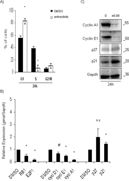
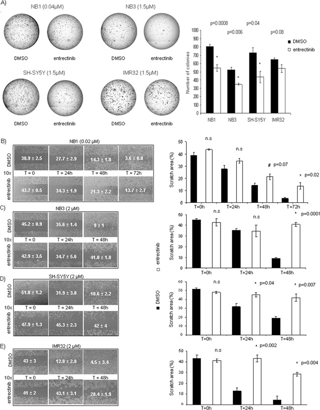
Neuroblastoma (NB) is a threatening childhood malignancy. Its prognosis is affected by several morphological, and biological characteristics, including the constitutive expression of ALK tyrosine kinase. In this study we examined the therapeutic potential of a novel ALK inhibitor, entrectinib, in obliterating NB tumor cells. Entrectinib showed the growth-inhibitory effects on NB cells with a 50% inhibitory concentration range of 0.03-5 μM. In the ALK-dependent cells, entrectinib mediated G1-arrest, which was associated with modified expression of multiple cell-cycle regulators. Down-regulation of Ki-67, and attenuated phosphorylation of ERK1/2, and STAT3, correlated with observed antiproliferative capacity of entrectinib. Initial cytostatic activity of entrectinib was followed by concentration-dependent apoptotic cell death, and Caspase-3 activation. However, we delineated a reduced sensitivity of ALK mutated NB cells to entrectinib, and demonstrated strong activation of autophagy in SH-SY5YF1174L NB cell line. Abrogation of autophagy by chloroquine increased significantly the toxicity of entrectinib, as confirmed by enhanced death rate, and PARP protein cleavage in SH-SY5YF1174L cells. In aggregate, our data show that entrectinib inhibits proliferation, and induces G1-arrest, and apoptosis in NB cells. We propose entrectinib for further consideration in treatment of NB, and recommend pharmacological inhibition of autophagy to be explored for a combined therapeutic approach in NB patients that might develop resistance to entrectinib.

Keywords: ALK inhibitors; autophagy; drug combination; neuroblastoma.


Conflict of interest statement

CONFLICTS OF INTEREST

The authors have no potential conflict of interest to declare.

Figures

Figure 1
Figure 1. Inhibition of cell viability, and growth after entrectinib treatment
A. NB cells were treated with increasing concentrations of entrectinib for 24h, 48h and 72h, and IC50 has been calculated for each cell line (NB1, NB3, SH-SY5Y and IMR32) for three time points. Concentrations used (μM) are indicated on X-axis. B. Trypan blue exclusion assay has been done to count the cell number in presence of three different concentration of entrectinib (2.5; 5; 7.5 μM). Data represent fold change of cell number calculated with respect to the number of cells present in the moment of administration of entrectinib, and expressed as mean ± SEM of 3 independent experiments. Time points are indicated on X-axis. C. Cell proliferation was measured by the means of Ki-67 mRNA expression 24h after addition of entrectinib (NB1 = 0.08 μM; NB3, SH-SY5Y, IMR32 = 2 μM). Results were presented as a relative expression calculated with respect to DMSO control (RQ = 1). Significant down-regulation of Ki-67 mRNA levels was evidenced for all cell lines. Single experiments were done in triplicates, and results presented for 3 separated treatments as mean ± SEM. Results were considered significant for *p ≤ 0.05.
Figure 2
Figure 2. Entrectinib modifies cell-cycle profile
A. Cell-cycle profile was evaluated by flow cytometry. Percentage of cells in each phase of cell-cycle is presented. An important accumulation of the NB1 cells in G1-phase was confirmed 24h post-treatment. B. qRT-PCR was performed for the evaluation of mRNA levels of the main cell-cycle regulators p21, p27, Cyclin A1, D1, E1, pRB1, and E2F1, after treatment with entrectinib (0.08 μM). Data were normalized for Gapdh internal control, and calculated with respect to the relative expression determined for DMSO control samples. Results were considered significant for p ≤ 0.05, and indicated with an asterisk (*). #p = 0.06. C. Expression of cell cycle regulatory proteins (p21, p27, Cyclin A1, and Cyclin E1) was validated by Western Blot 24h after treatment with entrectinib. Gapdh was used as a control of proper protein loading. Numbers indicate concentration of entrectinib (e; μM) used. D = DMSO.
Figure 3
Figure 3. Entrectinib impairs NB cell clonogenic competence, and motility
A. Representative images of clonogenic assays were shown for each cell line (left side). Cells were analyzed for the foci formation during the course of 14 days in the presence of either entrectinib or DMSO control vehicle. The figure shows the capacity of entrectinib to deteriorate the colony-forming cell. The number of colonies was counted by ImageJ software after coloration with MTT, and results shown as mean ± SEM (at right) of three independent experiments. Results were considered significant for *p < 0.05. Numbers within the brackets indicate the μM of entrectinib used in the experiment. B-E. Time-lapse microscopy reveals an inhibition of the motility of neuroblastoma cells upon entrectinib treatment (left side). For the wound healing assay, NB cells were treated with vehicle (DMSO control) and B) 0.02 μM (NB1) or C) 2 μM (NB3, SH-SY5Y, and IMR32) entrectinib, and monitored for 48-72h., The results are presented for each image (right side) as mean ± SEM of three separate experiments, and *p-value considered significant when ≤ 0.05. n.s.: not-significant; # - close to significance.
Figure 4
Figure 4. Entrectinib stimulates cell death in NB cell lines
A. The capability of entrectinib to induce Caspase-3 dependent death of NB cells was assessed applying flow cytometry technique. An increase in Caspase-3 activation was detected in different extents for diverse cell types 24h after treatment with increasing concentration of entrectinib. NB1amp was particularly sensitive to entrectinib, whereas SH-SY5YF1174L showed weaker sensitivity to the compound. B. Percentage of dead cells was measured by TUNEL assay, applying flow cytometry technique. The potential of entrectinib to impair cell viability was confirmed for all cell lines, for different concentration of entrectinib. The effects of entrectinib, and DMSO treated control samples are presented. Results were considered significant for *p ≤ 0.05. C. Activation of PARP protein, as a hallmark for death induction, was seen by Western blot. Cleavage of PARP protein was most evident for NB1 cell line. Gapdh was used as protein loading control. D = DMSO.
Figure 5
Figure 5. Entrectinib impacts ALK downstream protein pathway
Eventual change of ALK downstream pathway after treatment with entrectinib was validated by Western blot. Two different analyses were done: A. using entrectinib (e) IC50 defined for 48h, and B. using increasing concentration of entrectinib for 24h. Decreased activity of two principal downstream target of ALK protein, ERK1/2, and STAT3 can be seen by their reduced phosphorylation in either of immunoblots presented. Gapdh was used as a control of proper protein loading. Numbers beside letter (e) indicate concentration of entrectinib (μM) used. D = DMSO.
Figure 6
Figure 6. Entrectinib causes autophagy activation in ALK mutated NB cells
A. Immunoblot analyses of LC3 after treatment with IC50 (upper lane) or increasing concentration (lower lane) of entrectinib are presented. A significant processing of LC3 was seen for SH-SY5Y cells with ALK F1174L mutation. Gapdh was used as a protein loading control. B. Apoptosis (PARP), and autophagy (p62, and Beclin-1) related proteins were examined in SH-SY5Y cell line for several concentrations of entrectinib (e), and for 2 time points, 24h and 48h. Gapdh was used as a control of proper protein loading. Numbers beside letter (e) indicate concentration of entrectinib (μM) used in the experiments. Numbers alone indicate relative expression of the protein calculated by densitometry. D = DMSO. C. SH-SY5Y cells were transiently transfected with GFP-LC3 plasmid, and treated with entrectinib (5 μM) or DMSO vehicle for 24h. Fluorescence microscopic analysis was done afterwards to evaluate a distribution of GFP-LC3 within the cells. Cell nuclei were counterstained with DAPI (blue). Images were taken on a confocal microscope (100X immersion objective), equipped with a digital camera. D. Apoptosis (PARP), and autophagy (LC3, p62, and Beclin-1) related proteins were examined in NB3 cell line for increasing concentration of entrectinib. Gapdh was used as a control of proper protein loading. Initial numbers indicate concentration of entrectinib (e) in μM. Numbers under the blots indicate relative expression of the protein calculated by densitometry. D = DMSO.
Figure 7
Figure 7. Entrectinib activity in SH-SY5Y cells increases after abrogation of autophagy
A. Cells were pre-incubated with Chloroquine (CQ; 25 and 50 μM) for 1h, and entrectinib (e; 5 μM) was added in following for 24h. Whole cell lysates were used to evaluate the expression of PARP, p62, Beclin-1, and LC3 proteins for single or combined treatments. Gapdh was utilized as loading control. Numbers indicate concentration of compounds in μM used in the experiment. D = DMSO. B. Percentage of dead cells was measured by TUNEL assay, applying flow cytometry technique. The potential of entrectinib (e; 5 μM) to impair cell viability alone, or after pre-treatment with CQ (50 μM), was confirmed in SH-SY5Y cell line. The effects of treatments, and DMSO control samples are presented as mean ± SEM. Results were considered significant for *p ≤ 0.05. #p = 0.09. C. Tamoxifen (5 μM) and Rapamycin (10 μM) were used to stimulate autophagy in NB1 cells, whereas D) LY294002 (10 μM), and 3-MA (50 μM) were used as autophagy inhibitors in SH-SY5Y cells. Immunoblot analyses were performed to verify the levels of apoptosis- and autophagy-related proteins. Gapdh served as a loading control. Abbreviations: D, DMSO; e, entrectinib; T, Tamoxifen; Ra, Rapamycin; Ly, Ly294002; 3-MA, 3-Methyladenine. Numbers indicate concentration of entrectinib (μM) used for the experiments.
Figure 8
Figure 8. Autophagy activation is an early event in ALKF1174L mutated cells
A. Western blot analysis was done for the evaluation of LC3 processing, and hence, autophagy activation. NB1 and SH-SY5Y cells were treated shortly (6h) with entrectinib (e), using 0.04 μM, and 2.5 μM, respectively, and in following the expression of LC3-I, and LC3-II proteins was validated. We could confirm autophagy to be an early protective event in SH-SY5YF1174L cells, being activated after only 6h of treatment with entrectinib. On the contrary, in NB1amp cells, entrectinib showed an immediate apoptosis activation, as confirmed by cleavage of PARP protein, which has not been observed for SH-SY5Y cells. Gapdh was used as protein loading control. D = DMSO.
Figure 9
Figure 9. Effects of crizotinib on autophagy induction
Immunoblot analyses of autophagy (LC3, p62, and Beclin-1), and apoptosis (PARP) related proteins' expression after treatment with crizotinib are shown. Several concentrations of drug have been applied. Gapdh was used as a control of proper protein loading. Numbers beside letter (c) indicate concentration of crizotinib (μM) used in the experiments. Numbers under the blots indicate relative expression of the protein calculated by densitometry. D = DMSO.

Similar articles

Cited by

References

    1. SEER Program (National Cancer Institute (U.S.)) and National Cancer Institute (U.S.) 1999 Cancer incidence and survival among children and adolescents United States SEER Program, 1975-1995. Bethesda, Md: National Cancer Institute;
    1. Tonini GP, Nakagawara A, Berthold F. Towards a turning point of neuroblastoma therapy. Cancer Lett. 2012;326:128–134. - PubMed
    1. Maris JM. Recent advances in neuroblastoma. N Engl J Med. 2010;362:2202–2211. - PMC - PubMed
    1. Schwab M. MYCN in neuronal tumours. Cancer Lett. 2004;204:179–187. - PubMed
    1. Maris JM, Matthay KK. Molecular biology of neuroblastoma. J Clin Oncol. 1999;17:2264–2279. - PubMed

Publication types

MeSH terms