

Computer Tomography Imaging Findings of Abdominal Follicular Dendritic Cell Sarcoma: A Report of 5 Cases
- PMID: 26735543
- PMCID: PMC4706263
- DOI: 10.1097/MD.0000000000002404
Computer Tomography Imaging Findings of Abdominal Follicular Dendritic Cell Sarcoma: A Report of 5 Cases
Abstract
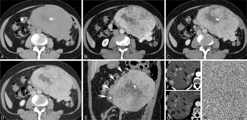
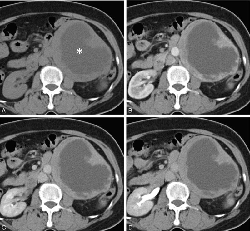
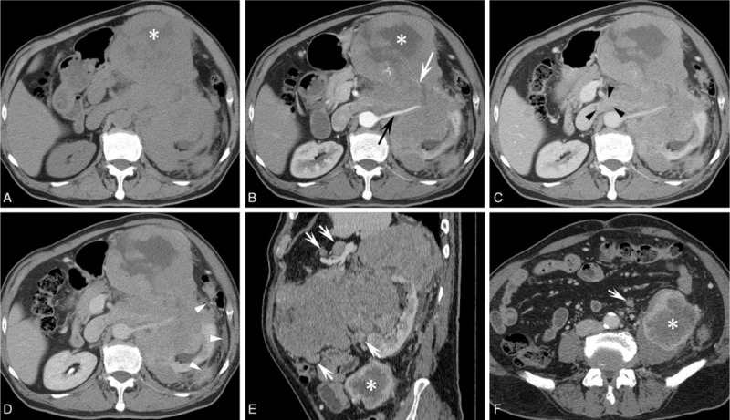
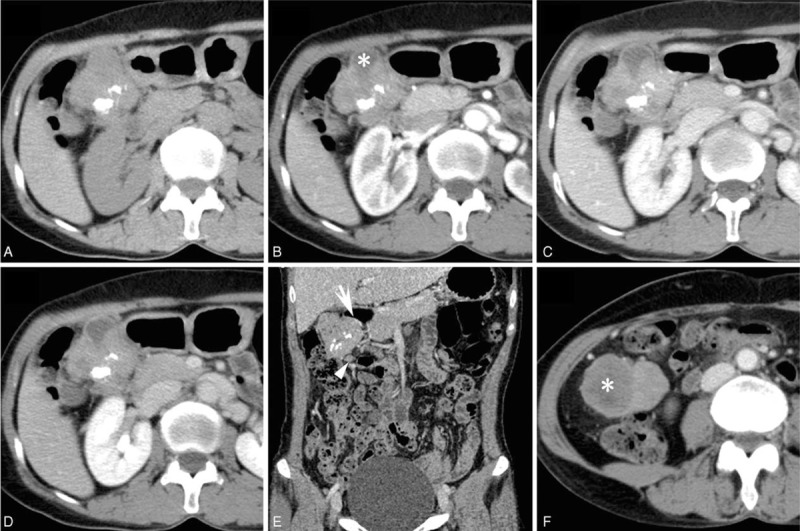
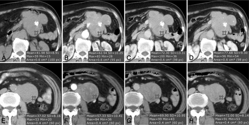
Follicular dendritic cell sarcoma (FDCS) is a neoplasm that arises from follicular dendritic cells. FDCSs originating in the abdomen are extremely rare. Clinically, they often mimic a wide variety of other abdominal tumors, and correct preoperative diagnosis is often a challenging task. To date, only scattered cases of abdominal FDCS have been reported and few data are available on their radiological features. Here we present the computer tomography imaging findings of 5 patients with surgically and pathologically demonstrated abdominal FDCS. An abdominal FDCS should be included in the differential diagnosis when single or multiple masses with relatively large size, well- or ill-defined borders, complex internal architecture with marked internal necrosis and/or focal calcification, and heterogeneous enhancement with "rapid wash-in and slow wash-out" or "progressive enhancement" enhancement patterns in the solid component are seen.
Conflict of interest statement
The authors have no conflicts of interest to disclose.
Figures
References
-
- Tew JG, Kosco MH, Burton GF, et al. Follicular dendritic cells as accessory cells. Immunol Rev 1990; 117:185–211. - PubMed
Publication types
MeSH terms
LinkOut - more resources
Full Text Sources
Other Literature Sources
