Pattern recognition with adaptive-thresholds for sleep spindle in high density EEG signals
- PMID: 26736332
- PMCID: PMC4888948
- DOI: 10.1109/EMBC.2015.7318432
Pattern recognition with adaptive-thresholds for sleep spindle in high density EEG signals
Abstract
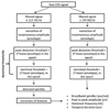
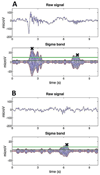
Medicine and Surgery, University of Pisa, via Savi 10, 56126, Pisa, Italy Sleep spindles are electroencephalographic oscillations peculiar of non-REM sleep, related to neuronal mechanisms underlying sleep restoration and learning consolidation. Based on their very singular morphology, sleep spindles can be visually recognized and detected, even though this approach can lead to significant mis-detections. For this reason, many efforts have been put in developing a reliable algorithm for spindle automatic detection, and a number of methods, based on different techniques, have been tested via visual validation. This work aims at improving current pattern recognition procedures for sleep spindles detection by taking into account their physiological sources of variability. We provide a method as a synthesis of the current state of art that, improving dynamic threshold adaptation, is able to follow modification of spindle characteristics as a function of sleep depth and inter-subjects variability. The algorithm has been applied to physiological data recorded by a high density EEG in order to perform a validation based on visual inspection and on evaluation of expected results from normal night sleep in healthy subjects.
Figures


References
-
- Wolpert EA. A manual of standardized terminology, techniques and scoring system for sleep stages of human subjects. Archives of General Psychiatry. 1969;20(2):246. - PubMed
-
- Menicucci D, Piarulli A, Allegrini P, Laurino M, Mastorci F, Sebastiani L, Bedini R, Gemignani A. Fragments of wake-like activity frame down-states of sleep slow oscillations in humans: New vistas for studying homeostatic processes during sleep. International Journal of Psychophysiology. 2013;89(2):151–157. - PubMed
-
- Steriade M. Grouping of brain rhythms in corticothalamic systems. Neuroscience. 2006;137(4):1087–1106. - PubMed
-
- De Gennaro L, Ferrara M. Sleep spindles: An overview. Sleep medicine reviews. 2003;7(5):423–440. - PubMed
Publication types
MeSH terms
Grants and funding
LinkOut - more resources
Full Text Sources
Research Materials
