Review
. 2016:56:339-60.
doi: 10.1146/annurev-pharmtox-010715-103617.

Targeting Prefrontal Cortical Systems for Drug Development: Potential Therapies for Cognitive Disorders

Affiliations
Review

Targeting Prefrontal Cortical Systems for Drug Development: Potential Therapies for Cognitive Disorders

Amy F T Arnsten et al. Annu Rev Pharmacol Toxicol. 2016.

Abstract

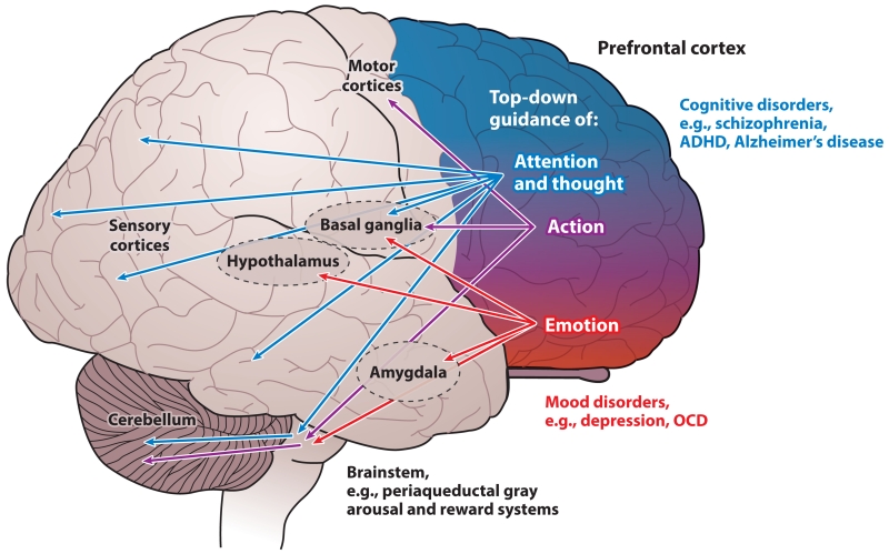
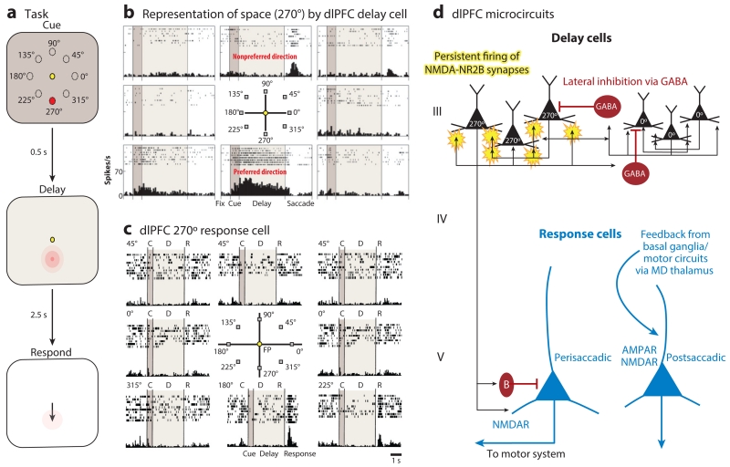
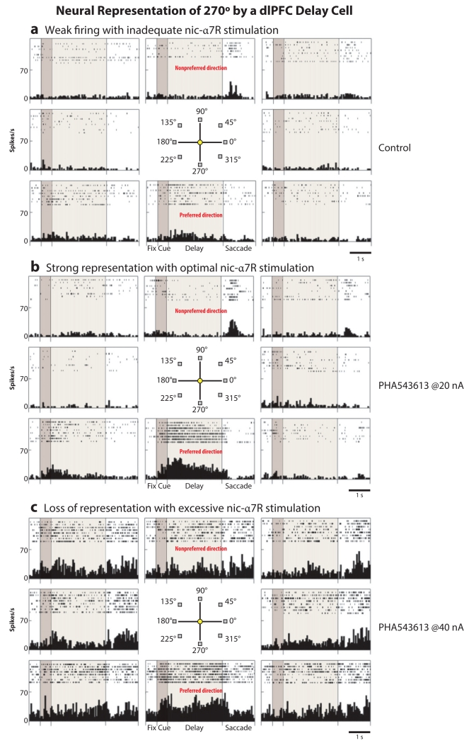
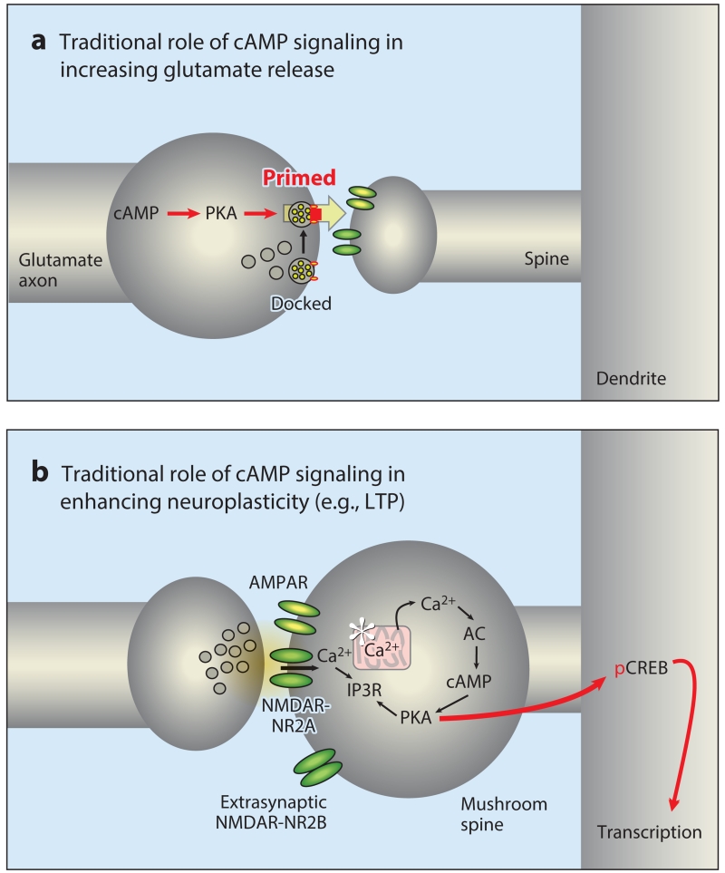
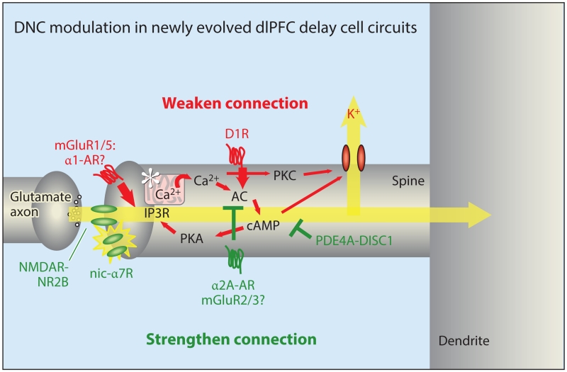
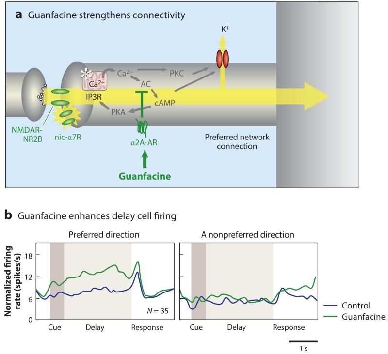
Medications to treat cognitive disorders are increasingly needed, yet researchers have had few successes in this challenging arena. Cognitive abilities in primates arise from highly evolved N-methyl-d-aspartate (NMDA) receptor circuits in layer III of the dorsolateral prefrontal cortex. These circuits have unique modulatory needs that can differ from the layer V neurons that predominate in rodents, but they offer multiple therapeutic targets. Cognitive improvement often requires low doses that enhance the pattern of information held in working memory, whereas higher doses can produce nonspecific changes that obscure information. Identifying appropriate doses for clinical trials may be helped by assessments in monkeys and by flexible, individualized dose designs. The use of guanfacine (Intuniv) for prefrontal cortical disorders was based on research in monkeys, supporting this approach. Coupling our knowledge of higher primate circuits with the powerful methods now available in drug design will help create effective treatments for cognitive disorders.

Keywords: Alzheimer's disease; acetylcholine; dopamine; norepinephrine; schizophrenia.


Figures

Figure 1
Figure 1
Topographic organization of PFC functions in primates. The primate PFC provides top-down guidance of attention and thought (blue), action (purple), and emotion (red) through its extensive projections. In general, the PFC is organized topographically, with dorsal and lateral regions regulating thought and attention and ventral and medial regions regulating emotion. This organization is reflected in the PFC projections through the basal ganglia via the dorsal and ventral striatum. There are also parallel projections to the cerebellum via the pontine nuclei. The human brain is lateralized, with the left hemisphere specialized for language and the right hemisphere specialized for inhibition of inappropriate thoughts, actions, and emotions. Dysfunction of the dorsal and lateral PFC is associated with cognitive disorders, whereas more ventral and medial areas are altered in affective disorders. Abbreviations: ADHD, attention deficit hyperactivity disorder; OCD, obsessive-compulsive disorder; PFC, prefrontal cortex.
Figure 2
Figure 2
The delay cells in layer III microcircuits in the dlPFC underlying spatial working memory. (a) Schematic illustration of the ODR task, in which a cue appears briefly (0.5 s) at one of eight locations (e.g., 270°) while the monkey fixates on a central spot. The location must be remembered over a delay period of several seconds until the fixation spot disappears and the monkey can move its eyes to the remembered location for a juice reward. The cued location constantly changes over hundreds of trials, requiring the constant updating of working memory. (b) An example of the firing patterns of a dlPFC delay cell representing the 270° location, the neuron’s preferred direction. This delay cell maintains firing across the delay epoch if the cue had appeared at 270° but not other locations. (c) An example of the firing patterns of a dlPFC response cell that is providing feedback during the eye movement to 270°. Response cells are often inhibited during the delay epoch. (d) The microcircuitry in the primate dlPFC thought to underlie working memory. Microcircuits underlying delay cell firing are thought to reside in deep layer III, the layer that expanded most in primate evolution. Clusters of pyramidal cells with similar preferred directions excite each other to maintain persistent firing across the delay period in the absence of sensory stimulation. This requires glutamate stimulation of NMDAR-NR2B. In contrast, the spatial specificity is refined by lateral inhibition from parvalbumin-containing GABAergic interneurons. In contrast, response cells are thought to reside in layer V. Perisaccadic response cells fire leading up to the motor response, and postsaccadic response cells are thought to carry the corollary discharge feedback that a response has occurred. The postsaccadic response cells are influenced by both NMDAR and AMPAR stimulation. Delay cells likely inhibit response cells during the delay epoch via an inhibitory interneuron. Abbreviations: AMPAR, α-amino-3-hydroxy-5-methyl-4-isoxazolepropionic acid receptor; dlPFC, dorsolateral prefrontal cortex; MD, mediodorsal; NMDA, N-methyl-d-aspartate; NMDAR, NMDA receptor; ODR, oculomotor delayed response.
Figure 3
Figure 3
Neural representation of 270° by a dlPFC delay cell. Low, but not high, doses of a nic-α7R agonist enhance the neural representation of visual space by dlPFC delay cells. (a) A dlPFC delay cell has weak, delay-related firing under control conditions, with subtle representation of 270°. (b) Iontophoresis of a low dose of the nic-α7R agonist PHA543613 (at 20 nA) enhances the representation of 270° by increasing persistent firing across the delay period only in trials in which the monkey is remembering 270°. (c) A higher dose of PHA543613 (at 40 nA) increases persistent firing for all directions, thus eroding the information held in working memory stores. Thus, low doses are often essential to be effective cognitive enhancers. Abbreviations: dlPFC, dorsolateral prefrontal cortex; nic-α7R, nicotinic α7 receptor.
Figure 4
Figure 4
The traditional roles of cAMP-PKA signaling in modulating neurotransmission and neuroplasticity. (a) cAMP-PKA signaling can enhance transmitter release (e.g., glutamate release) from the presynaptic terminal by priming vesicles for release. (b) cAMP-PKA signaling is needed for late-stage LTP in hippocampal neurons. These synapses generally contain AMPAR and NMDAR-NR2A subunits, whereas NMDAR-NR2Bs are found extrasynaptically. This process can involve internal calcium release from a spine apparatus in some synapses. Sufficient activation of PKA can lead to phosphorylation of CREB and transcriptional changes that can lead to spine enlargement (an immature, thin learning spine becomes a mushroom spine) and/or enlargement and stabilization of the PSD. Abbreviations: AC, adenylyl cyclase; AMPAR, α-amino-3-hydroxy-5-methyl-4-isoxazolepropionic acid receptor; cAMP, cyclic adenosine monophosphate; CREB, cAMP response element–binding protein; IP3R, inositol 1,4,5-trisphosphate receptor; LTP, long-term potentiation; NMDA, N-methyl-d-aspartate; NMDAR, NMDA receptor; pCREB, phosphorylated CREB; PKA, protein kinase A; PSD, postsynaptic density.
Figure 5
Figure 5
DNC modulation in newly evolved dlPFC delay cell circuits. DNC in a mature, long, thin spine in layer III of the primate dlPFC alters synapse strength rapidly and reversibly to coordinate cognitive and arousal states. In contrast to traditional synapses, NMDAR-NR2Bs are localized exclusively in the PSD and are not extrasynaptic. These synapses have only a subtle AMPAR component, and the permissive excitation needed for NMDAR opening is instead mediated by cholinergic stimulation of nic-α7R (and possibly muscarinic M1R). Mechanisms that increase feedforward calcium-cAMP-PKA signaling (red, many engaged by stress) weaken synaptic connections by opening nearby K+ channels (HCN and KCNQ) on the spine head, neck, or both. In contrast, inhibiting feedforward calcium-cAMP-PKA signaling (green) strengthens connectivity and enhances delay cell firing. Loss of inhibition, e.g., through genetic insults to DISC1, may contribute to spine loss and impaired dlPFC function. The unique modulation of layer III dlPFC pyramidal cell synapses provides strategies for therapeutic targets for cognitive disorders. Abbreviations: AC, adenylyl cyclase; AMPAR, α-amino-3-hydroxy-5-methyl-4-isoxazolepropionic acid receptor; AR, adrenergic receptor; cAMP, cyclic adenosine monophosphate; D1R, dopamine D1 receptor; DISC1, Disrupted In SChizophrenia 1; dlPFC, dorsolateral prefrontal cortex; DNC, dynamic network connectivity; HCN, hyperpolarization-activated cyclic nucleotide gated channels; IP3R, inositol 1,4,5-trisphosphate receptor; KCNQ, potassium channel, voltage-gated, KQT-like subfamily; M1R, muscarinic M1 receptor; mGluR, metabotropic glutamate receptor; nic-α7R, nicotinic α7 receptor; NMDA, N-methyl-d-aspartate; NMDAR, NMDA receptor; PDE4A, phosphodiesterase 4A; PKA/PKC, protein kinase A/C.
Figure 6
Figure 6
The α2A-AR agonist guanfacine strengthens dlPFC network connections and enhances dlPFC network firing. (a) A schematic illustration of guanfacine’s actions in the primate dlPFC, engaging postsynaptic α2A-ARs to inhibit feedforward cAMP-calcium-K+ channel signaling and strengthen NMDAR connections. (b) Guanfacine increases the firing of delay cells for their preferred direction. The normalized mean firing rate of 35 delay cells under control conditions (blue) and following iontophoresis of guanfacine (green) are shown, as are the neurons’ preferred direction and their nonpreferred direction opposite to the preferred direction. Guanfacine improves a variety of PFC cognitive functions and is now in widespread clinical use. Abbreviations: AC, adenylyl cyclase; AR, adrenergic receptor; cAMP, cyclic adenosine monophosphate; dlPFC, dorsolateral PFC; IP3R, inositol 1,4,5-trisphosphate receptor; nic-α7R, nicotinic α7 receptor; NMDA, N-methyl-d-aspartate; NMDAR, NMDA receptor; PFC, prefrontal cortex; PKA/PKC, protein kinase A/C.

References

    1. Lewis DA, Campbell MJ, Terry RD, Morrison JH. Laminar and regional distributions of neurofibrillary tangles and neuritic plaques in Alzheimer’s disease: a quantitative study of visual and auditory cortices. J. Neurosci. 1987;7:1799–808. - PMC - PubMed
    1. Bussière T, Giannakopoulos P, Bouras C, Perl DP, Morrison JH, Hof PR. Progressive degeneration of nonphosphorylated neurofilament protein-enriched pyramidal neurons predicts cognitive impairment in Alzheimer’s disease: stereologic analysis of prefrontal cortex area 9. J. Comp. Neurol. 2003;463:281–302. - PubMed
    1. Lewis DA, Gonzalez-Burgos GR. Pathophysiologically based treatment interventions in schizophrenia. Nat. Med. 2006;12:1016–22. - PubMed
    1. Arion D, Corradi JP, Tang S, Datta D, Boothe F, et al. Distinctive transcriptome alterations of prefrontal pyramidal neurons in schizophrenia and schizoaffective disorder. Mol. Psychiatry. 2015;20:1397–405. - PMC - PubMed
    1. Arnsten AFT, Wang M, Paspalas CD. Neuromodulation of thought: flexibilities and vulnerabilities in prefrontal cortical network synapses. Neuron. 2012;76:223–39. - PMC - PubMed

Publication types

LinkOut - more resources