

A systems approach to hemostasis: 4. How hemostatic thrombi limit the loss of plasma-borne molecules from the microvasculature
- PMID: 26738537
- PMCID: PMC4807424
- DOI: 10.1182/blood-2015-09-672188
A systems approach to hemostasis: 4. How hemostatic thrombi limit the loss of plasma-borne molecules from the microvasculature
Abstract
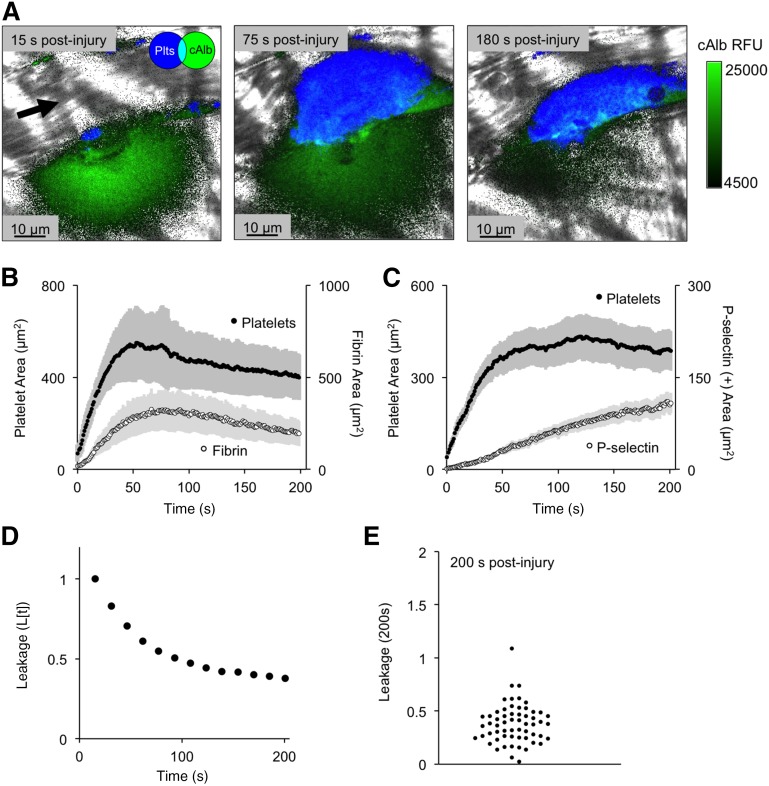
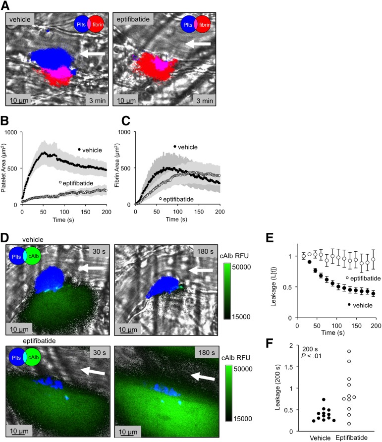
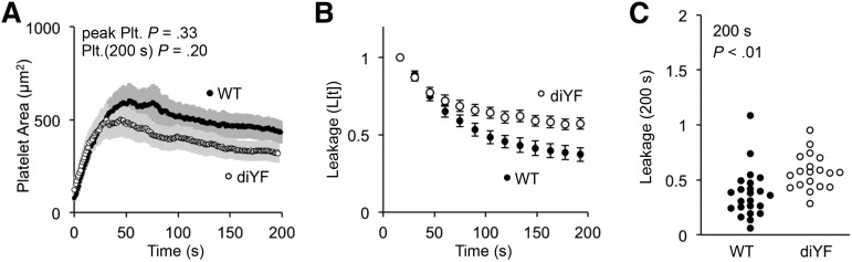
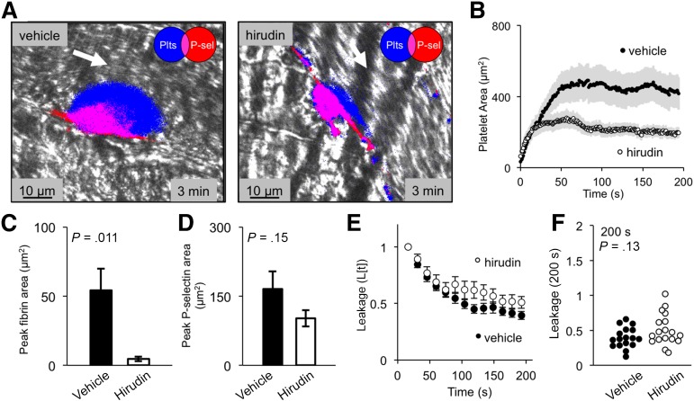
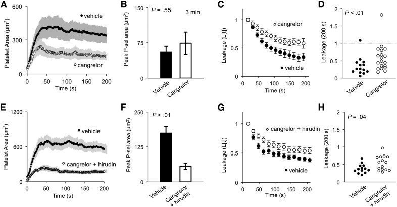
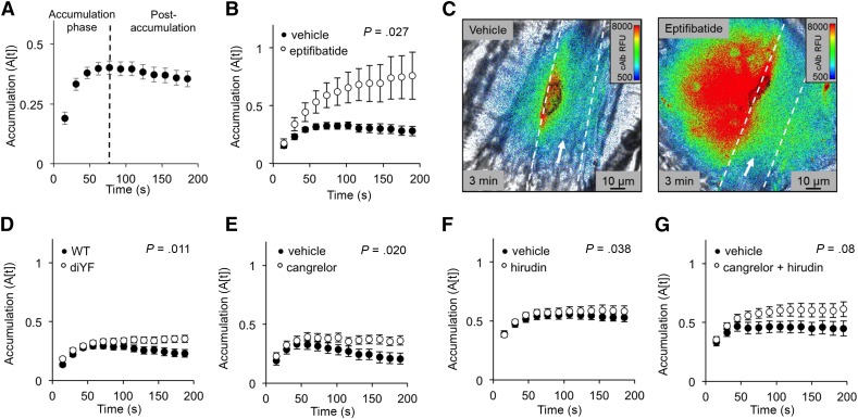
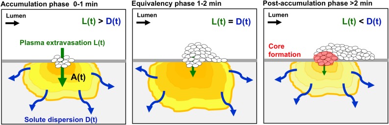
Previous studies have shown that hemostatic thrombi formed in response to penetrating injuries have a core of densely packed, fibrin-associated platelets overlaid by a shell of less-activated, loosely packed platelets. Here we asked, first, how the diverse elements of this structure combine to stem the loss of plasma-borne molecules and, second, whether antiplatelet agents and anticoagulants that perturb thrombus structure affect the re-establishment of a tight vascular seal. The studies combined high-resolution intravital microscopy with a photo-activatable fluorescent albumin marker to simultaneously track thrombus formation and protein transport following injuries to mouse cremaster muscle venules. The results show that protein loss persists after red cell loss has ceased. Blocking platelet deposition with an αIIbβ3antagonist delays vessel sealing and increases extravascular protein accumulation, as does either inhibiting adenosine 5'-diphosphate (ADP) P2Y12receptors or reducing integrin-dependent signaling and retraction. In contrast, sealing was unaffected by introducing hirudin to block fibrin accumulation or a Gi2α gain-of-function mutation to expand the thrombus shell. Collectively, these observations describe a novel approach for studying vessel sealing after injury in real time in vivo and show that (1) the core/shell architecture previously observed in arterioles also occurs in venules, (2) plasma leakage persists well beyond red cell escape and mature thrombus formation, (3) the most critical events for limiting plasma extravasation are the stable accumulation of platelets, ADP-dependent signaling, and the emergence of a densely packed core, not the accumulation of fibrin, and (4) drugs that affect platelet accumulation and packing can delay vessel sealing, permitting protein escape to continue.
© 2016 by The American Society of Hematology.
Figures
Comment in
-
Platelets stop us leaking.Blood. 2016 Mar 24;127(12):1528-9. doi: 10.1182/blood-2016-01-692186. Blood. 2016. PMID: 27013214
Similar articles
-
Platelet packing density is an independent regulator of the hemostatic response to injury.J Thromb Haemost. 2018 May;16(5):973-983. doi: 10.1111/jth.13986. Epub 2018 Apr 2. J Thromb Haemost. 2018. PMID: 29488682 Free PMC article.
-
Hierarchical organization of the hemostatic response to penetrating injuries in the mouse macrovasculature.J Thromb Haemost. 2017 Mar;15(3):526-537. doi: 10.1111/jth.13600. Epub 2017 Feb 6. J Thromb Haemost. 2017. PMID: 27992950 Free PMC article.
-
Hierarchical organization in the hemostatic response and its relationship to the platelet-signaling network.Blood. 2013 Mar 7;121(10):1875-85. doi: 10.1182/blood-2012-09-457739. Epub 2013 Jan 9. Blood. 2013. PMID: 23303817 Free PMC article.
-
Interactions of platelets, blood-borne tissue factor, and fibrin during arteriolar thrombus formation in vivo.Microcirculation. 2005 Apr-May;12(3):301-11. doi: 10.1080/10739680590925682. Microcirculation. 2005. PMID: 15814438 Review.
-
Platelets and coagulation in thrombus formation: aberrations in the Scott syndrome.Thromb Res. 2016 May;141 Suppl 2:S12-6. doi: 10.1016/S0049-3848(16)30355-3. Thromb Res. 2016. PMID: 27207414 Review.
Cited by
-
Platelet packing density is an independent regulator of the hemostatic response to injury.J Thromb Haemost. 2018 May;16(5):973-983. doi: 10.1111/jth.13986. Epub 2018 Apr 2. J Thromb Haemost. 2018. PMID: 29488682 Free PMC article.
-
GLS-409, an Antagonist of Both P2Y1 and P2Y12, Potently Inhibits Canine Coronary Artery Thrombosis and Reversibly Inhibits Human Platelet Activation.Sci Rep. 2018 Sep 28;8(1):14529. doi: 10.1038/s41598-018-32797-1. Sci Rep. 2018. PMID: 30266987 Free PMC article.
-
Impact of Tissue Factor Localization on Blood Clot Structure and Resistance under Venous Shear.Biophys J. 2018 Feb 27;114(4):978-991. doi: 10.1016/j.bpj.2017.12.034. Biophys J. 2018. PMID: 29490257 Free PMC article.
-
Visualizing thrombosis to improve thrombus resolution.Res Pract Thromb Haemost. 2021 Jan 6;5(1):38-50. doi: 10.1002/rth2.12469. eCollection 2021 Jan. Res Pract Thromb Haemost. 2021. PMID: 33537528 Free PMC article.
-
12(S)-HETrE, a 12-Lipoxygenase Oxylipin of Dihomo-γ-Linolenic Acid, Inhibits Thrombosis via Gαs Signaling in Platelets.Arterioscler Thromb Vasc Biol. 2016 Oct;36(10):2068-77. doi: 10.1161/ATVBAHA.116.308050. Epub 2016 Jul 28. Arterioscler Thromb Vasc Biol. 2016. PMID: 27470510 Free PMC article.
References
-
- Bellido-Martín L, Chen V, Jasuja R, Furie B, Furie BC. Imaging fibrin formation and platelet and endothelial cell activation in vivo. Thromb Haemost. 2011;105(5):776–782. - PubMed
-
- van Gestel MA, Heemskerk JW, Slaaf DW, et al. Real-time detection of activation patterns in individual platelets during thromboembolism in vivo: differences between thrombus growth and embolus formation. J Vasc Res. 2002;39(6):534–543. - PubMed
-
- Hechler B, Nonne C, Eckly A, et al. Arterial thrombosis: relevance of a model with two levels of severity assessed by histologic, ultrastructural and functional characterization. J Thromb Haemost. 2010;8(1):173–184. - PubMed
-
- Hayashi T, Mogami H, Murakami Y, et al. Real-time analysis of platelet aggregation and procoagulant activity during thrombus formation in vivo. Pflugers Arch. 2008;456(6):1239–1251. - PubMed
Publication types
MeSH terms
Substances
Grants and funding
LinkOut - more resources
Full Text Sources
Other Literature Sources
Medical
Molecular Biology Databases
