. 2016 Jan 6:13:6.
doi: 10.1186/s12974-015-0474-6.

CXCL5 signaling is a shared pathway of neuroinflammation and blood-brain barrier injury contributing to white matter injury in the immature brain

Affiliations

CXCL5 signaling is a shared pathway of neuroinflammation and blood-brain barrier injury contributing to white matter injury in the immature brain

Lin-Yu Wang et al. J Neuroinflammation. .

Abstract

Background: In very preterm infants, white matter injury is a prominent brain injury, and hypoxic ischemia (HI) and infection are the two primary pathogenic factors of this injury. Microglia and microvascular endothelial cells closely interact; therefore, a common signaling pathway may cause neuroinflammation and blood-brain barrier (BBB) damage after injury to the immature brain. CXC chemokine ligand 5 (CXCL5) is produced in inflammatory and endothelial cells by various organs in response to insults. CXCL5 levels markedly increased in the amniotic cavity in response to intrauterine infection and preterm birth in clinical studies. The objective of this study is to determine whether CXCL5 signaling is a shared pathway of neuroinflammation and BBB injury that contributes to white matter injury in the immature brain.

Methods: Postpartum day 2 (P2) rat pups received lipopolysaccharide (LPS) followed by 90-min HI. Immunohistochemical analyses were performed to determine microglial activation, neutrophil infiltration, BBB damage, and myelin basic protein and glial fibrillary acidic protein expression. Immunofluorescence experiments were performed to determine the cellular distribution of CXCL5. Pharmacological tests were performed to inhibit or enhance CXCL5 activity.

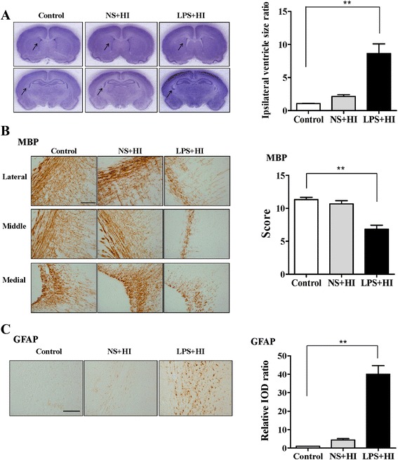
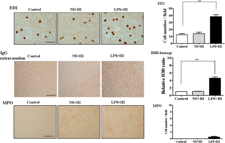
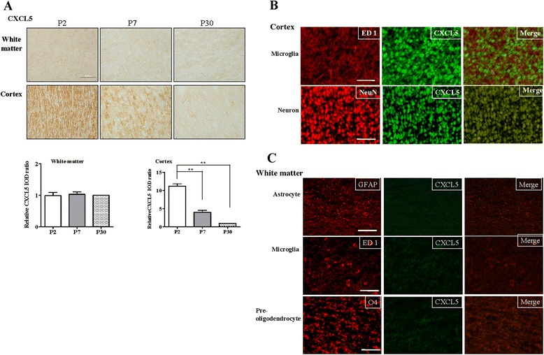
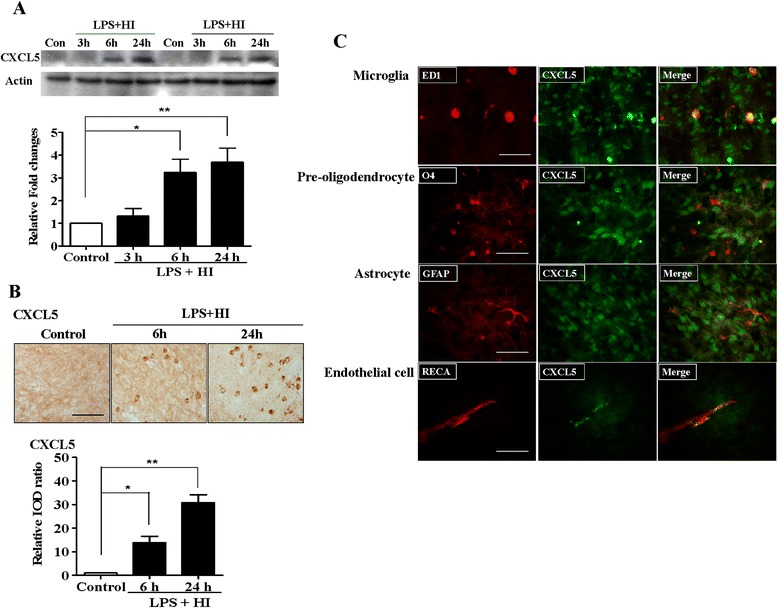
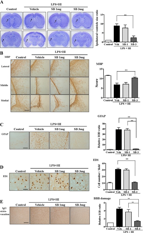
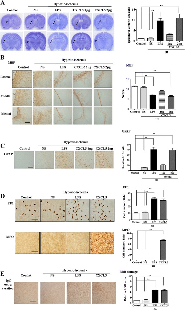
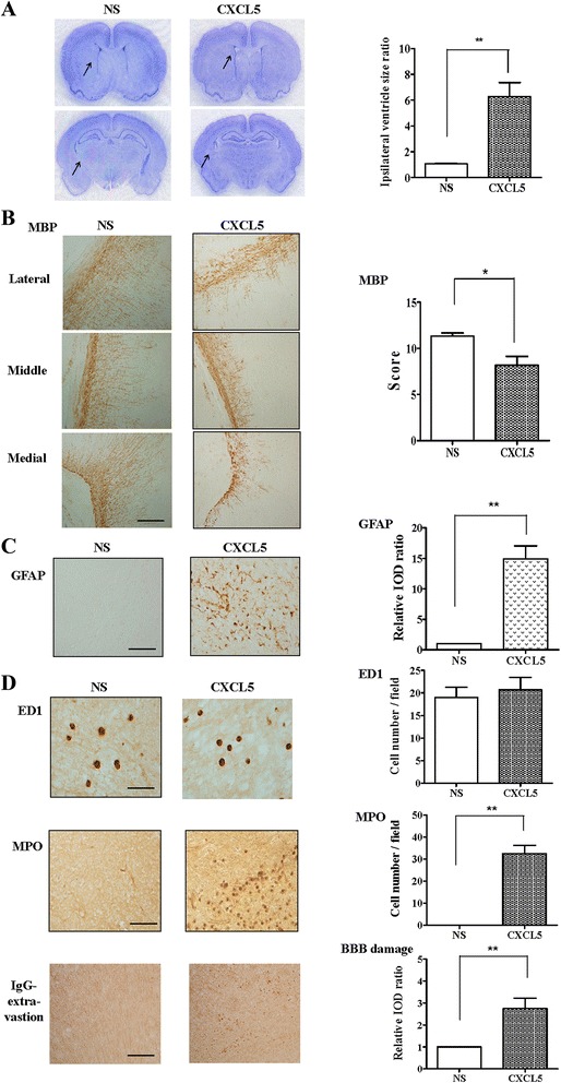
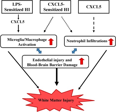
Results: On P2, predominant increases in microglial activation and BBB damage were observed 24 h after LPS-sensitized HI induction, and white matter injury (decreased myelination and increased astrogliosis) was observed on P12 compared with controls. Immunohistochemical analyses revealed increased CXCL5 expression in the white matter 6 and 24 h after insult. Immunofluorescence experiments revealed upregulated CXCL5 expression in the activated microglia and endothelial cells 24 h after insult. CXCL5 inhibition by SB225002, a selective nonpeptide inhibitor of CXCR2, significantly attenuated microglial activation and BBB damage, increased myelination, and reduced astrogliosis in the white matter after LPS-sensitized HI. In addition, CXCL5-sensitized HI or CXCL5 alone significantly induced BBB damage and white matter injury in association with different neuroinflammation mechanisms. CXCL5-sensitized HI-induced microglial activation and neutrophil infiltration, whereas CXCL5 alone predominately caused neutrophil infiltration.

Conclusions: CXCL5 is a potential biomarker for white matter injury in preterm infants. Pharmacological blockade of CXCL5 signaling that attenuates dysregulated neuroinflammation can be used a therapeutic strategy against white matter injury in the immature brain.


Figures

Fig. 1
Fig. 1
LPS-sensitized HI caused selective white matter injury. P2 rat pups received LPS (0.05 mg/kg) or NS 3 h before 90 min of HI. Neuropathological tests were performed on P12 (a, left panel). Nissl staining revealed no significant injury in the gray matter of the control (n = 6), NS + HI (n = 6), and LPS + HI (n = 6) groups. Upper panel: brain section at the level of striatum (0.26 mm posterior to the bregma) and lower panel: the section at the level of dorsal hippocampus (3.14 mm posterior to the bregma). The LPS + HI group had significantly higher ipsilateral ventricular size ratios (a, right panel), lower myelination (MBP) (b), and higher astrogliosis (GFAP) (c) in the white matter than those of the control group. Scale bar = 100 μm for MBP and GFAP. Values are means ± SEMs. **p < 0.01. Values are means ± SEMs. NS, normal saline; LPS, lipopolysaccharide; HI, hypoxic ischemia; MBP, myelin basic protein; GFAP, glial fibrillary acidic protein
Fig. 2
Fig. 2
LPS-sensitized HI induced microglial activation and BBB damage. Neuroinflammation and BBB damage were assessed 24 h after insult. The LPS + HI group had a significantly higher number of ED1(+) activated microglia (upper panel) and significantly greater IgG extravasation suggesting BBB damage (middle panel) but fewer MPO(+) neutrophils (lower panel) in the white matter than did the control group (n = 6 in each group). BBB, blood–brain barrier. Scale bar = 50 μm (ED1) and = 100 μm (IgG and MPO). Values are means ± SEMs, **p < 0.01
Fig. 3
Fig. 3
CXCL5 expression in the normal developing rat brain. a CXCL5 immunohistochemical analysis demonstrated that P7 (n = 4) and, particularly, P2 pups (n = 5) had significantly higher CXCL5 expression in the cerebral cortex than that of P30 rats (n = 4). Values are means ± SEMs. The P2 pups had significantly higher CXCL5 expression in the cerebral cortex than that of the P7 pups and P30 rats. CXCL5 expression was low in the white matter, and no significant difference was observed between the three groups. b Immunofluorescence staining in the P2 pups revealed that the most CXCL5-positive cells in the cortex were neurons (NeuN) rather than microglia (ED1), and CXCL5 expression was absent in all white matter cells. c Scale bar = 50 μm
Fig. 4
Fig. 4
CXCL5 upregulation in the white matter after LPS-sensitized HI. a Western blots of the white matter revealed significant increases in CXCL5 levels at 6 and 24 h after LPS + HI (n = 4 in each study group). b Immunohistochemical analyses revealed that the LPS + HI group had significantly increased CXCL5 immunoreactivity in the white matter at 6 h and, particularly, 24 h after HI compared with the control group (n = 6 in each study group). Scale bar = 50 μm. Values are means ± SEMs. c Immunofluorescence analysis of the white matter 24 h after LPS + HI revealed that CXCL5 was upregulated mainly in activated microglia (ED1) and vascular endothelial cells (RECA) and not in astrocytes (GFAP) or preoligodendrocytes (O4). Scale bar = 50 μm. *p < 0.05, **p < 0.01
Fig. 5
Fig. 5
Pharmacological inhibition of CXCR2 significantly attenuated microglial activation and BBB damage and protected against white matter injury after LPS-sensitized HI. A selective nonpeptide inhibitor of CXCR2, SB22 5002, was used to examine the role of the CXCL5–CXCR2 pathway in white matter injury after LPS-sensitized HI. After LPS + HI on P2, the SB-3 group (n = 9), but not the SB-1 group (n = 7), had significantly reduced ipsilateral ventricular size ratios (a), increased myelination (MBP) (b), and reduced astrogliosis (GFAP) (c) in the white matter on P12 compared with the vehicle-treated pups (n = 9). Scale bar = 100 μm. The SB-3 group (n = 9), but not the SB-1 group (n = 7), had a significantly reduced number of activated microglia (ED1) (d) and significantly lower BBB damage (IgG extravasation) (e) in the white matter compared with the vehicle group (n = 9) at 24 h after HI. Scale bar = 50 μm (ED1) and 100 μm (IgG); values are means ± SEMs, **p < 0.01
Fig. 6
Fig. 6
CXCL5-sensitized HI induced microglial activation, neutrophil infiltration, and BBB damage and caused white matter injury. Intracerebroventricular infusion of recombinant CXCL5 (1 or 2 μg) or NS followed by HI on P2 showed that the 2-μg CXCL5 + HI group (n = 6), but not the 1-μg CXCL5 + HI group (n = 6), had significantly higher ipsilateral ventricle size ratios (a), lower myelination (MBP) (b), and higher astrogliosis (GFAP) (c) in the white matter compared with the NS (n = 6) and the control groups (n = 6) on P12. Scale bar = 100 μm. At 24 h after HI, the 2-μg CXCL5 + HI (n = 6) and LPS + HI groups (n = 6) had a significantly higher number of activated microglia (ED1) (d) and BBB damage (e) in the white matter compared with the NS (n = 6) and control groups (n = 6). The 2-μg CXCL5 + HI group, but not the LPS + HI group, had significantly increased neutrophil infiltrations (MPO) (d) in the white matter. Scale bar = 50 μm (ED1) and 100 μm (IgG and MPO); values are means ± SEMs, **p < 0.01
Fig. 7
Fig. 7
CXCL5 alone increased neutrophil infiltration and BBB damage and caused white matter injury. Intracerebroventricular infusion of recombinant CXCL5 (2 μg) or NS on P2 revealed that the CXCL5 group (n = 6) had significantly higher ipsilateral ventricle size ratios (a), reduced myelination (MBP) (b), and increased astrogliosis (GFAP) (c) in the white matter on P12 compared with the vehicle group (n = 6). Scale bar = 100 μm. (d) At 24 h after injection, the NS (n = 6) and CXCL5 groups (n = 6) showed no detectable ED1(+) activated microglia in the white matter. By contrast, the CXCL5 group had a significantly higher number of MPO(+) neutrophils and significantly greater BBB damage compared with the vehicle group. Scale bar = 50 μm (ED1) and 100 μm (IgG and MPO); values are means ± SEMs, *p < 0.05, **p < 0.01
Fig. 8
Fig. 8
Diagram showing that the proposed CXCL5-mediated signaling pathway, triggered by LPS-sensitized HI, CXCL5-sensitized HI, or CXCL5 alone, plays a crucial role in neuroinflammation and BBB disruption and subsequent white matter injury in the immature brain. Neuroinflammation and BBB damage are two mutually potentiating mechanisms leading to sustained neuroinflammation and BBB disruption in white matter injury of the developing brain. White matter injury can be induced by different neuroinflammatory mechanisms: predominant microglial activation by LPS-sensitized HI, microglial activation and neutrophil infiltration by CXCL5-sensitized HI, and predominant neutrophil infiltration by CXCL5 alone

References

    1. Raines EW, Ferri N. Thematic review series: the immune system and atherogenesis. Cytokines affecting endothelial and smooth muscle cells in vascular disease. J Lipid Res. 2005;46:1081–1092. doi: 10.1194/jlr.R500004-JLR200. - DOI - PubMed
    1. Charo IF, Ransohoff RM. The many roles of chemokines and chemokine receptors in inflammation. New Engl J Med. 2006;354:610–621. doi: 10.1056/NEJMra052723. - DOI - PubMed
    1. Sepuru KM, Poluri KM, Rajarathnam K. Solution structure of CXCL5—a novel chemokine and adipokine implicated in inflammation and obesity. PLoS One. 2014;9:e93228. doi: 10.1371/journal.pone.0093228. - DOI - PMC - PubMed
    1. Walz A, Schmutz P, Mueller C, Schnyder-Candrian S. Regulation and function of the CXC chemokine ENA-78 in monocytes and its role in disease. J Leukoc Biol. 1997;62:604–611. - PubMed
    1. Rovai LE, Herschman HR, Smith JB. The murine neutrophil-chemoattractant chemokines LIX, KC, and MIP-2 have distinct induction kinetics, tissue distributions, and tissue-specific sensitivities to glucocorticoid regulation in endotoxemia. J Leukoc Biol. 1998;64:494–502. - PubMed

Publication types

MeSH terms