

Clinical diagnostics and therapy monitoring in the congenital disorders of glycosylation
- PMID: 26739145
- PMCID: PMC4891361
- DOI: 10.1007/s10719-015-9639-x
Clinical diagnostics and therapy monitoring in the congenital disorders of glycosylation
Abstract
Abnormal protein glycosylation is observed in many common disorders like cancer, inflammation, Alzheimer's disease and diabetes. However, the actual use of this information in clinical diagnostics is still very limited. Information is usually derived from analysis of total serum N-glycan profiling methods, whereas the current use of glycoprotein biomarkers in the clinical setting is commonly based on protein levels. It can be envisioned that combining protein levels and their glycan isoforms would increase specificity for early diagnosis and therapy monitoring. To establish diagnostic assays, based on the mass spectrometric analysis of protein-specific glycosylation abnormalities, still many technical improvements have to be made. In addition, clinical validation is equally important as well as an understanding of the genetic and environmental factors that determine the protein-specific glycosylation abnormalities. Important lessons can be learned from the group of monogenic disorders in the glycosylation pathway, the Congenital Disorders of Glycosylation (CDG). Now that more and more genetic defects are being unraveled, we start to learn how genetic factors influence glycomics profiles of individual and total serum proteins. Although only in its initial stages, such studies suggest the importance to establish diagnostic assays for protein-specific glycosylation profiling, and the need to look beyond the single glycoprotein diagnostic test. Here, we review progress in and lessons from genetic disease, and review the increasing opportunities of mass spectrometry to analyze protein glycosylation in the clinical diagnostic setting. Furthermore, we will discuss the possibilities to expand current CDG diagnostics and how this can be used to approach glycoprotein biomarkers for more common diseases.
Keywords: Congenital disorders of glycosylation; Glycomics; Protein-specific glycosylation; Transferrin.
Figures
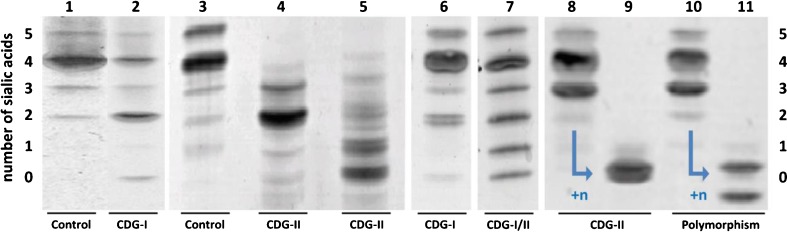

 ) and define the CDG-I-component of this phenotype. The yellow arrow (
 ) demonstrates the absence of galactose on one of the truncated glycans, which define the CDG type-II component. The number of sialic acids is indicated above each structure, and the insets at the right show respective IEF results
References
Publication types
MeSH terms
Substances
LinkOut - more resources
Full Text Sources
Other Literature Sources
