Review
. 2016 Jan 6:15:4.
doi: 10.1186/s12943-015-0481-3.

Imaging metabolic heterogeneity in cancer

Affiliations
Review

Imaging metabolic heterogeneity in cancer

Debanti Sengupta et al. Mol Cancer. .

Abstract

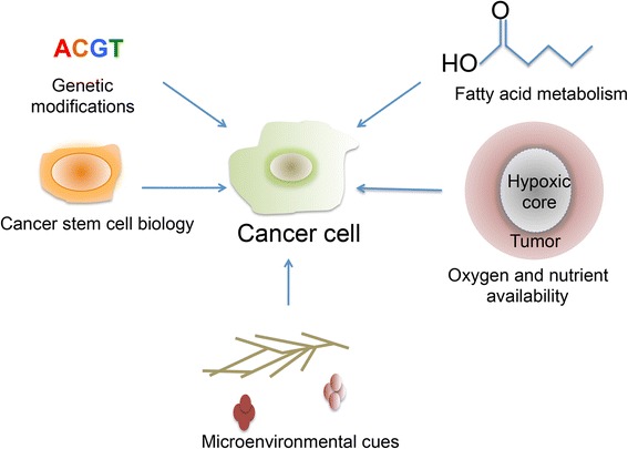
As our knowledge of cancer metabolism has increased, it has become apparent that cancer metabolic processes are extremely heterogeneous. The reasons behind this heterogeneity include genetic diversity, the existence of multiple and redundant metabolic pathways, altered microenvironmental conditions, and so on. As a result, methods in the clinic and beyond have been developed in order to image and study tumor metabolism in the in vivo and in vitro regimes. Both regimes provide unique advantages and challenges, and may be used to provide a picture of tumor metabolic heterogeneity that is spatially and temporally comprehensive. Taken together, these methods may hold the key to appropriate cancer diagnoses and treatments in the future.


Figures

Fig. 1
Fig. 1
A schematic of factors that influence cancer metabolic heterogeneity
Fig. 2
Fig. 2
a PET/CT images comparing [11C] -acetate and FDG in a prostate cancer patient. This research was originally published in the Journal of Nuclear Medicine. Oyama N, Akino H, Kanamaru H, Suzuki Y, Muramoto S, Yonekura Y, Sadato N, Yamamoto K, Okada K: 11C-acetate PET imaging of prostate cancer. J Nucl Med 2002, 43:181-186. © by the Society of Nuclear Medicine and Molecular Imaging, Inc [58]. b Hyperpolarized MR imaging of hyperpolarized [13C] pyruvate and its conversion to [13C] alanine and [13C] lactate in vivo. Adapted by permission from the American Association for Cancer Research: Golman K, Lerche M, Pehrson R, Ardenkjaer-Larsen JH: Metabolic imaging by hyperpolarized 13C magnetic resonance imaging for in vivo tumor diagnosis. Cancer Research 2006, 66:10855-10860 [58]
Fig. 3
Fig. 3
a Endogenous fluorescence images of NADH and FAD lifetimes in organoids derived from triple negative breast cancer (TNBC) and HER2+ tumors. Adapted by permission from the American Association for Cancer Research: Walsh AJ, Cook RS, Sanders ME, Aurisicchio L, Ciliberto G, Arteaga CL, Skala MC: Quantitative optical imaging of primary tumor organoid metabolism predicts drug response in breast cancer. Cancer Res 2014, 74:5184-5194 [124]. b Radioluminescence microscopy of single MDA-MB-231 human breast cancer cells that have taken up FDG

References

    1. Hsu PP, Sabatini DM. Cancer cell metabolism: Warburg and beyond. Cell. 2008;134:703–707. doi: 10.1016/j.cell.2008.08.021. - DOI - PubMed
    1. Garber K. Energy deregulation: licensing tumors to grow. Science. 2006;312:1158–1159. doi: 10.1126/science.312.5777.1158. - DOI - PubMed
    1. Cairns RA, Harris IS, Mak TW. Regulation of cancer cell metabolism. Nat Rev Cancer. 2011;11:85–95. doi: 10.1038/nrc2981. - DOI - PubMed
    1. Gatenby RA, Gillies RJ. Why do cancers have high aerobic glycolysis? Nat Rev Cancer. 2004;4:891–899. doi: 10.1038/nrc1478. - DOI - PubMed
    1. Gillies RJ, Robey I, Gatenby RA. Causes and consequences of increased glucose metabolism of cancers. J Nucl Med. 2008;49:24S–42S. doi: 10.2967/jnumed.107.047258. - DOI - PubMed