. 2016 Jan 19;113(3):656-61.
doi: 10.1073/pnas.1509834113. Epub 2016 Jan 6.

Extracardiac septum transversum/proepicardial endothelial cells pattern embryonic coronary arterio-venous connections

Affiliations

Extracardiac septum transversum/proepicardial endothelial cells pattern embryonic coronary arterio-venous connections

Elena Cano et al. Proc Natl Acad Sci U S A. .

Abstract

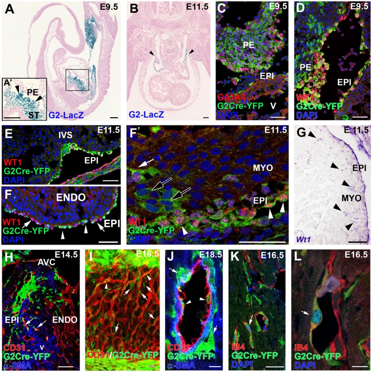
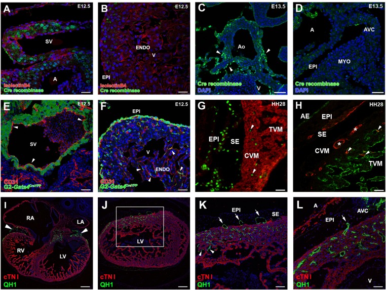
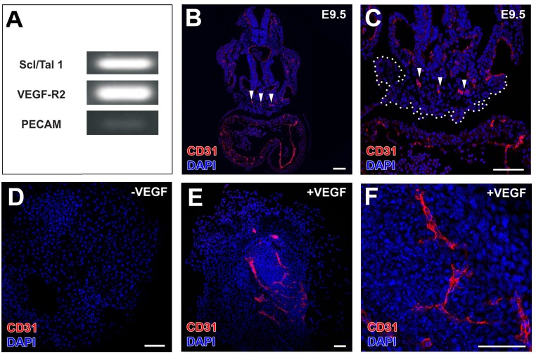
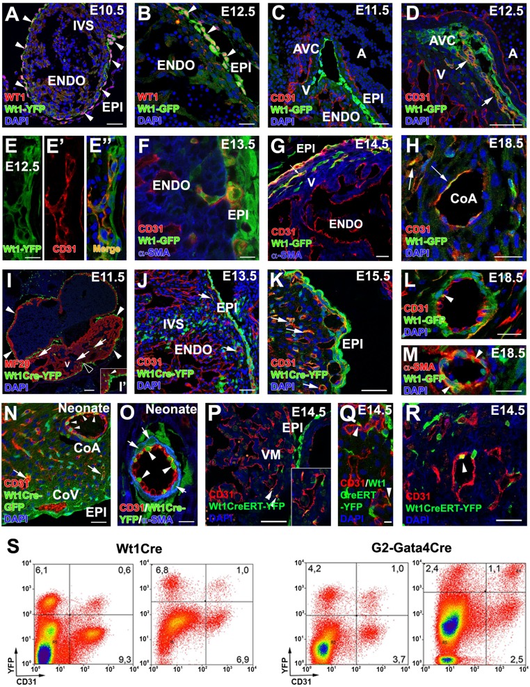
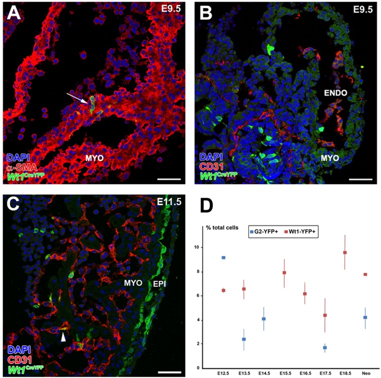
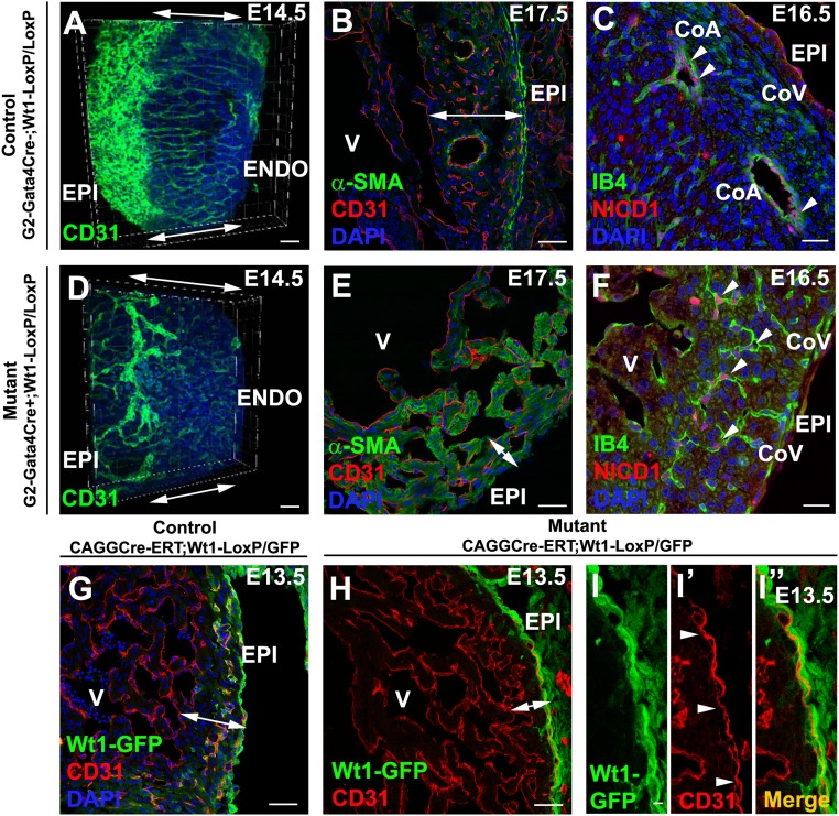
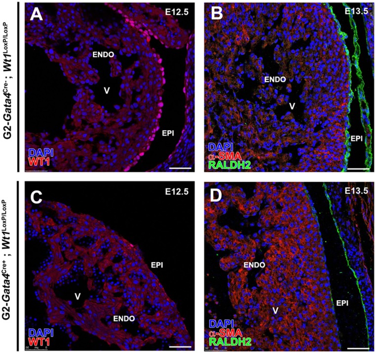
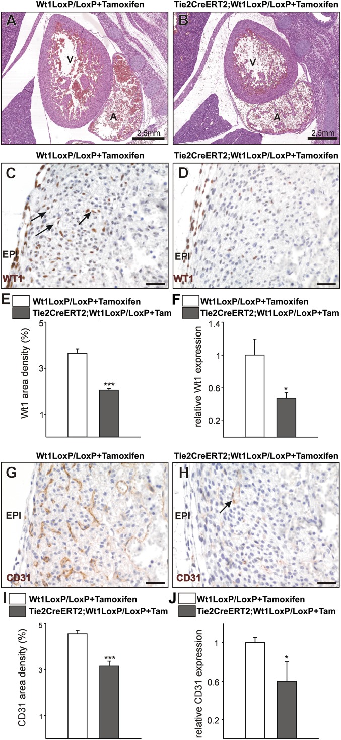
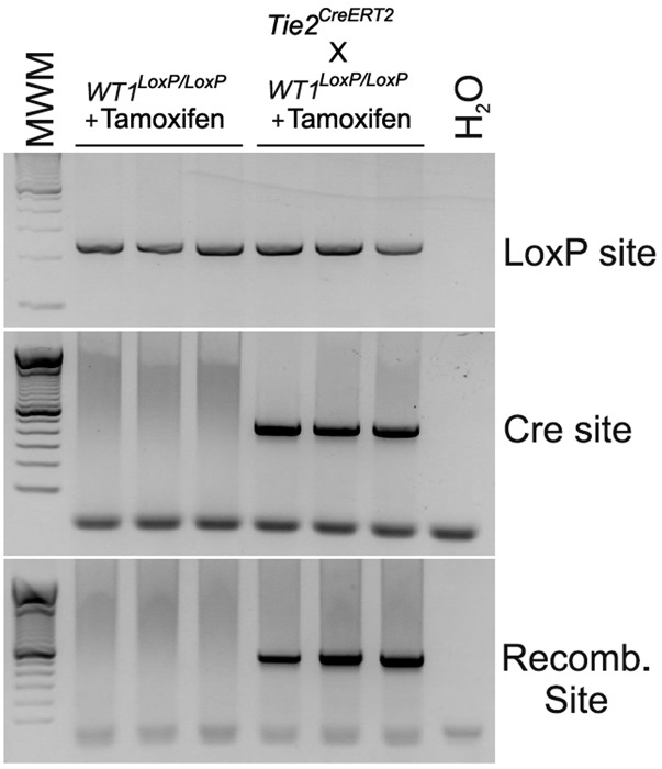
Recent reports suggest that mammalian embryonic coronary endothelium (CoE) originates from the sinus venosus and ventricular endocardium. However, the contribution of extracardiac cells to CoE is thought to be minor and nonsignificant for coronary formation. Using classic (Wt1(Cre)) and previously undescribed (G2-Gata4(Cre)) transgenic mouse models for the study of coronary vascular development, we show that extracardiac septum transversum/proepicardium (ST/PE)-derived endothelial cells are required for the formation of ventricular coronary arterio-venous vascular connections. Our results indicate that at least 20% of embryonic coronary arterial and capillary endothelial cells derive from the ST/PE compartment. Moreover, we show that conditional deletion of the ST/PE lineage-specific Wilms' tumor suppressor gene (Wt1) in the ST/PE of G2-Gata4(Cre) mice and in the endothelium of Tie2(Cre) mice disrupts embryonic coronary transmural patterning, leading to embryonic death. Taken together, our results demonstrate that ST/PE-derived endothelial cells contribute significantly to and are required for proper coronary vascular morphogenesis.

Keywords: Gata4; Wt1; coronary endothelium; proepicardium; septum transversum.


Conflict of interest statement

The authors declare no conflict of interest.

Figures

Fig. 1.
Fig. 1.
ST/PE G2-Gata4 cells throughout cardiac development. (A and B) G2-Gata4LacZ mice show reporter activity in the septum transversum (including the PE) at E9.5 (A) and inflow myocardium at E11.5 (B, arrowheads). (C and D) Immunohistochemistry of G2-Gata4CreYFP+ samples illustrates the expression of GATA4 (C) and WT1 (D) proteins in G2-Gata4CreYFP+ cells at E9.5 PE. (EF′) G2-Gata4CreYFP mice show an increasing number of G2-Gata4CreYFP+ cells from the developing epicardium in subepicardial and intramyocardial areas between stages E10.5 and E14.5. The epicardium comprises WT1+/G2-Gata4+ (arrowheads) and WT1+/G2-Gata4 cells (F, arrows). A few EPDCs retain WT1 expression transiently (F and F′, white arrow), but other EPDCs do not (F′, black arrows). (G) Wt1 gene expression is conspicuous in the epicardium but is restricted to a few EPDCs (arrowheads). (H) Progressive expansion of EPDCs through the myocardial walls at E14.5–18.5 parallels G2-Gata4CreYFP+ incorporation in developing coronary blood vessels (arrows). (I and J) 3D reconstructions (I) and tissue section analysis (J) of the developing coronary vasculature allow perivascular cells (arrows in I and J) to be distinguished from G2-Gata4CreYFP+ CoE cells (arrowheads in I and J). (K and L) Identification of active Notch1 signaling by Notch1 intracellular domain (N1ICD) nuclear localization confirms the arterial nature of these vessels (arrows). A, atrium; AVC, atrio–ventricular canal; ENDO, endocardium; EPI, epicardium; IVS, interventricular septum; MYO, myocardium; PE, proepicardium; ST, septum transversum; V, ventricle. (Scale bars: 100 µm in A and B; 50 µm in CH; 40 µm in I; 10 µm in J; 25 µm in K; 5 µm in L.)
Fig. S1.
Fig. S1.
Origin and vascularizing properties of ST/PE-derived endothelial cells. (AD) Cre recombinase expression in G2CreYFP embryos is evident in the inflow myocardium at E12.5 (A) and in the aortic walls at E13.5 (C, arrowheads) but not in the epicardium at E12.5 (B) or epicardium or myocardial layers at E13.5 (D). (E and F) In G2CreYFP embryos, the sinus venosus endocardium is YFP (arrowheads in E). Some G2-Gata4CreYFP+/CD31+ cells also can be found incorporated in the ventricular endocardium (arrowheads in F). (G) Incorporation of PE-derived endothelial cells in the ventricular endocardium also occurs in quail-to-chick PE chimeric transplantations. PE derivatives, which express the quail pan-nuclear marker QCPN (green), invade the myosin-expressing ventricular walls (MF20+, red); some of these cells are incorporate in the ventricular endocardium (arrowheads). (H) The endothelial nature of these quail PE-derived cells is confirmed further by their expression of the QH1 endothelial marker (red). After FITC-conjugated Lens culinaris agglutinin counterstaining of the endocardium (green), PE-derived CoE cells (asterisks) can be distinguished easily from those that fused with the endocardium (arrowheads). (IL) Transplantation of posterior pericardiac mesenchyme without PE tissue (homologous to the mammalian ST) extensively vascularizes the embryonic myocardium (green QH1+ cells in I and J; arrowheads in I mark the accumulation of these cells at the atrioventricular canal). The area boxed in J is magnified in K to show endothelial invasion of the myocardial layers (red, cTNI+) from the subepicardial space (arrows). The same phenomenon is also evident at the atrioventricular canal (arrow in L). Some ST-derived endothelial cells also are incorporated in the endocardium (arrowheads in K). A, atrium; AE, atrial endocardium; AVC, atrioventricular canal; CVM, compact ventricular endocardium; ENDO, endocardium; EPI, epicardium; LA, left atrium; LV, left ventricle; RA, right atrium; RV, right ventricle; SE, subepicardium; SV, sinus venosus; TVM, trabeculated ventricular myocardium; V, ventricle. (Scale bars: 25 µm in A and B; 50 µm in CH and L; 150 µm in I and J; 75 µm in K.)
Fig. S2.
Fig. S2.
The E9.5 PE holds an intrinsic vascular potential. (A) Semiquantitative RT-PCRs confirm that E9.5 PE cells express vascular progenitor (Scl/Tal1/Vegfr-2) and endothelial (CD31) markers. (B and C) Low CD31 expression in PE is in accordance with the accumulation of differentiated CD31+ CoE cells in the dorsal aspect of the PE (arrowheads in B and C; the whole PE is marked by a dotted line in C). (DF) In vitro-cultured E9.5 PE differentiate into CD31+ CoE cells only in the presence of VEGF (compare E and F with D). (Scale bars: 100 µm.)
Fig. 2.
Fig. 2.
Wt1-expressing cells and their progeny contribute to CoE. (A and B) WT1 protein is ubiquitously expressed in epicardial cells and EPDCs at E10.5–E12.5, extensively overlapping with Wt1 promoter-driven GFP expression (arrowheads). (CG) Reporter expression in Wt1GFP embryos can be observed in the CD31+ subepicardial (EE”) and intramyocardial (F and G) coronary vasculature at E12.5–E14.5 (arrows in D and G), but not at earlier developmental stages (C). (H) A few Wt1GFP+/CD31+ cells are still found at perinatal stages (arrows). (I, I′, and J) Early Wt1CreYFP+ cells form the epicardium (arrowheads in I; the area marked with a black arrowhead is magnified in I′) and the first EPDCs (arrows in J). At E11.5 many Wt1CreYFP+ epicardial cells show morphological EMT features (arrowhead in I′). (J and K) The lineage reporter colocalizes with the vascular marker CD31 in the subepicardial and intramyocardial coronary plexus between E13.5 (J) and E15.5 (K) (arrows). (LO) Perinatal (L and M) and neonatal (N and O) hearts show Wt1CreYFP+ cells incorporated in the CoE (arrowheads in L and O) and CoSM layers of large CoA (arrowheads in M and arrows in O) as well as in capillaries (arrow in N). (PR) Wt1CreERT2;Rosa26-YFP embryos induced with tamoxifen at E9.5 show YFP+ cell incorporation in coronary vessels at E.14.5 (arrowheads). (S) Representative cytograms of dissociated ventricles from midgestation embryos and neonates. Numbers indicate percentages of total events. Both the Wt1CreYFP+ and G2CreYFP+ populations include CD31+ in cells. A, atrium; AVC, atrio–ventricular canal; CoA, coronary artery; CoV, coronary vein; ENDO, endocardium; EPI, epicardium; IVS, interventricular septum; V, ventricle. (Scale bars: 100 µm in I; 50 µm in A, C, D, J, M, N, and P; 25 µm in B, FH, K, and L; 25 µm in R; 10 µm in EE′′ and O; 5 µm in Q.)
Fig. S3.
Fig. S3.
Wt1 lineage-derived cardiomyocytes in the E9.0 mouse heart. (A and B) A minority of Wt1CreYFP+ cells can be found in the developing heart before or during PE cell transference to the myocardial surface (E9.5). These Wt1CreYFP+ cells express myocardial markers (α-SMA; arrow in A) but not vascular markers (CD31+) (B). (C) At early epicardial stages (E10.5–11.5) some rare endocardial Wt1CreYFP+/CD31+ cells could be recorded (arrowhead). (D) The total percentage of cardiac Wt1CreYFP+ and G2CreYFP+ cells throughout the course of development is shown. ENDO, endocardium; EPI, epicardium; MYO, myocardium. (Scale bars: 50 µm.)
Fig. 3.
Fig. 3.
G2-Gata4 and conditional systemic Wt1 deletion disrupt CoA formation. (AF) G2-Gata4Cre;Wt1LoxP/LoxP embryonic CoE structures are dysmorphic and fail to contact the endocardium (compare double-headed arrows in A and D and compare Movie S3 with Movie S4). The mutants show dramatic reduction of compact ventricular myocardium thickness (compare double-headed arrows in B and E). N1ICD immunohistochemistry identifies dysmorphic intramyocardial vessels as developing CoA (arrowheads in C and F), whereas subepicardial vascular structures are N1ICD (prospective CoV). (G and H) Tamoxifen-induced (E10.5) systemic Wt1 deletion sharply reduces the number of developing intramyocardial blood vessels. (II′′) Subepicardial blood vessels (arrowheads) form normally. CoA, coronary artery; CoV, coronary vein; ENDO, endocardium; EPI, epicardium; V, ventricle. (Scale bars: 50 µm in A and B; 25 µm in C; 50 µm in D, E, GI; 25 µm in F.)
Fig. S4.
Fig. S4.
G2Cre-driven Wt1 deletion. Effective deletion of Wt1 in the G2-Gata4 lineage is confirmed by the absence of WT1 protein (compare A and C) and by the decrease in the RALDH2 enzyme, a known direct Wt1 target (compare B and D). ENDO, endocardium; EPI, epicardium; V, ventricle. (Scale bars: 50 µm.)
Fig. 4.
Fig. 4.
Endothelial Wt1 expression is required for coronary vessel formation. (A and B) H&E-stained sections from control Wt1LoxP/LoxP+Tamoxifen (A) and mutant TieCreERT2;Wt1LoxP/LoxP+Tamoxifen (B) E16.5 embryos. (CF) The WT1+ cell contribution to coronary vessels (arrows in C) is reduced in the mutants (D and E), and Wt1 gene expression in E16.5 mutant hearts is reduced (F). (G and H) E16.5 mutant embryonic hearts show a decrease in compact ventricular wall coronary CD31+ cells (arrow in H; compare with G). (I and J) Quantification of the area occupied by CD31+ cells (I) and CD31 gene expression (J) in E16.5 hearts. A, atrium; EPI, epicardium; V, ventricle. (Scale bars: 50 µm.) Data are mean ± SEM; *P < 0.05, ***P < 0.001.
Fig. S5.
Fig. S5.
Tie2-CreERT-driven Wt1 deletion. Western blotting shows the specificity of tamoxifen-induced recombination in Wt1LoxP/LoxP mice versus Tie2CreERT2;Wt1LoxP/LoxP mutant mice.
Fig. 5.
Fig. 5.
A model of the EPDC contribution to the CoE. ST/PE-derived endothelial cells (green) give rise to the epicardium and EPDCs (AC) and are incorporated in intramyocardial CoA and capillary endothelium (D). Major transmural endothelial cell flows are indicated by arrow color; the arrow size estimates the frequency of the events. A, atrium; A-V, arterio–venous; CoA, coronary arteries; CoV: coronary veins; EPI: epicardium; PE: proepicardium; ST, septum transversum; SV, sinus venosus; V, ventricle.

References

    1. Pérez-Pomares JM, de la Pompa JL. Signaling during epicardium and coronary vessel development. Circ Res. 2011;109(12):1429–1442. - PubMed
    1. Red-Horse K, Ueno H, Weissman IL, Krasnow MA. Coronary arteries form by developmental reprogramming of venous cells. Nature. 2010;464(7288):549–553. - PMC - PubMed
    1. Wu B, et al. Endocardial cells form the coronary arteries by angiogenesis through myocardial-endocardial VEGF signaling. Cell. 2012;151(5):1083–1096. - PMC - PubMed
    1. Tian X, et al. Vessel formation. De novo formation of a distinct coronary vascular population in neonatal heart. Science. 2014;345(6192):90–94. - PMC - PubMed
    1. Tian X, Pu WT, Zhou B. Cellular origin and developmental program of coronary angiogenesis. Circ Res. 2015;116(3):515–530. - PMC - PubMed

Publication types

MeSH terms

LinkOut - more resources