Review
. 2016 Mar;468(3):455-73.
doi: 10.1007/s00424-015-1777-2. Epub 2016 Jan 6.

A Pore Idea: the ion conduction pathway of TMEM16/ANO proteins is composed partly of lipid

Affiliations
Review

A Pore Idea: the ion conduction pathway of TMEM16/ANO proteins is composed partly of lipid

Jarred M Whitlock et al. Pflugers Arch. 2016 Mar.

Abstract

Since their first descriptions, ion channels have been conceived as proteinaceous conduits that facilitate the passage of ionic cargo between segregated environments. This concept is reinforced by crystallographic structures of cation channels depicting ion conductance pathways completely lined by protein. Although lipids are sometimes present in fenestrations near the pore or may be involved in channel gating, there is little or no evidence that lipids inhabit the ion conduction pathway. Indeed, the presence of lipid acyl chains in the conductance pathway would curse the design of the channel's aqueous pore. Here, we make a speculative proposal that anion channels in the TMEM16/ANO superfamily have ion conductance pathways composed partly of lipids. Our reasoning is based on the idea that TMEM16 ion channels evolved from a kind of lipid transporter that scrambles lipids between leaflets of the membrane bilayer and the modeled structural similarity between TMEM16 lipid scramblases and TMEM16 anion channels. This novel view of the TMEM16 pore offers explanation for the biophysical and pharmacological oddness of TMEM16A. We build upon the recent X-ray structure of nhTMEM16 and develop models of both TMEM16 ion channels and lipid scramblases to bolster our proposal. It is our hope that this model of the TMEM16 pore will foster innovative investigation into TMEM16 function.

Keywords: Anoctamin; Calcium; Chloride channel; Phospholipid scrambling; Protein-lipid interactions; TMEM16.


Figures

Fig. 1
Fig. 1
The TMEM16/Anoctamin (ANO) family tree. a A phylogenetic tree generated from 1650 TMEM16 sequences in Uniprot. Non-redundant sequences were aligned by MUSCLE [63] and columns containing >50 % gaps were removed with TrimAl [20]. Phylogenetic trees were constructed by CLCBIO Main Workbench 6.9 using Kimura Neighbor-Joining. The fungal TMEM16 proteins afTMEM16 and nhTMEM16 are indicated. b A subset of vertebrate TMEM16 proteins identified by Uniprot were assembled and curated to remove splice variants and duplicate sequences. The sequences were truncated by deleting (~50) variable N-terminal amino acids. Trees were displayed using Dendroscope (http://dendroscope.org/). Percent identity and (similarity) refer to human proteins compared to human TMEM16A. Brief description of known disease relevance follows sequence alignments
Fig. 2
Fig. 2
Phospholipid scrambling is a ubiquitous cell signaling process. Left: Phospholipids are asymmetrically distributed between the two leaflets of the plasma membrane. PtdCho and sphingomyelin (open blue circles) are concentrated in the outer leaflet while PtdEtn and PtdSer (solid red circles) are concentrated in the inner leaflet. Right: Phospholipid scrambling stimulated by elevation of cytosolic Ca2+ or by apoptotic caspase activation results lipid mixing that exposes PtdSer and PtdEtn on the external leaflet. PtdSer and PtdEt exposure results in assembly of various macromolecular complexes (ligand binding) and membrane trafficking events associated with cell fusion and production of microvesicles [29, 42, 45, 50, 51, 118]
Fig. 3
Fig. 3
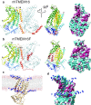
Phospholipid scrambling by TMEM16 proteins. a Crystal structure of nhTMEM16 [17] (4WIS) and b a homology model of TMEM16F based on the nhTMEM16 structure using Phyre2 [54]. One monomer is colored rainbow (blue is N-terminus, red is C-terminus) and the other is grey- light blue. Helices are numbered. Left panels: dimer viewed from the plane of the membrane. Middle panels: one monomer rotated 90° around the y-axis. The scrambling (SCRD) domain in TMEM16F in B is colored firebrick red. Right panels: molecular surfaces of the same view as the middle panel. Cyan = hydrophilic (−4.5, Kyte-Doolittle scale). Magenta = hydrophobic (4.5). c Molecular dynamics simulation of interaction of lipids with nhTMEM16 (http://sbcb.bioch.ox.ac.uk/memprotmd/beta/protein/pdbid/4WIS). A bilayer-embedded model was produced from the nhTMEM16 crystal structure through the MemProtMD protocol [104]. Lipids are shown in wire representation and the nitrogens of the choline headgroups of PtdCho molecules near the protein are shown as blue spheres. PtdCho molecules can be seen in the hydrophilic furrow and clustering near the SCRD. d Molecular surface of a close-up view of the hydrophilic furrow. The orientation is the same as c. Only the N of the PtdCho choline head groups is shown. Images were created using UCSF Chimera v. 1.10
Fig. 4
Fig. 4
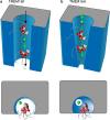
Hypothesis for evolution of a Cl channel from a phospholipid scramblase. As discussed in the text, we believe that PLS mediated by TMEM16F is associated with leakage of ions through the lipid scrambling pathway (the hydrophilic furrow) between the protein and the scrambling lipid head groups. Structural changes in the phospholipid scrambling pathway during evolution may have produced a Cl selective channel by decreasing phospholipid mobility in the furrow while still retaining the association of the head groups with the protein as a structural component. In this case, ions would still be capable of flowing between the lipid head groups and the protein. Ionic selectivity would be determined by both the protein and cognate phospholipids. a Cartoon of the scrambling furrow showing TMEM16F scramblase. Top panel is viewed in perspective from the plane of the membrane. Bottom panel is a view down the furrow from the extracellular space. Two PtdCho molecules are shown moving through furrow along with a Cl ion. The phospholipids are shown with their acyl tails in stick representation projecting into the hydrophobic bilayer. The polar head group atoms are shown as spheres with the atoms colored by element (grey = carbon, red = oxygen, orange = phosphorous). b Cartoon of TMEM16A. Two PtdCho molecules are lodged in the furrow because it is too narrow for them to move. However, Cl ions can slip between the lipid head groups and the protein. The effective diameter of the “pore” is imagined as smaller in TMEM16A than TMEM16F as seen in the lower panels. Although only 2 phospholipid molecules are shown, we calculate that the furrow is filled with 4–5 phospholipid molecules creating a monolayer that joins the outer and inner leaflets
Fig. 5
Fig. 5
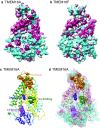
The TMEM16A furrow likely forms the conduction pathway for Cl. Molecular surface of homology models of a TMEM16A and b TMEM16F. Homology models were made from nhTMEM16 structure (4WIS) using Phyre2 [54]. Cyan = hydrophilic (−4.5, Kyte-Doolittle scale). Magenta = hydrophobic (4.5). Note hydrophobic region at the base of the furrow in TMEM16A that is hydrophilic in TMEM16F. c Functional residues of TMEM16A. Homology model of TMEM16A with functional amino acids identified by mutagenesis shown as spheres. Orange: vestibule [86, 129]. Blue: selectivity [126]. Green: Ca2+ binding [126, 129]. Magenta: gating modifier EEEEEAVK [125]. Transmembrane helices are colored as in Fig. 3. d Model of TMEM16A with a superimposed surface colored by hydrophobicity showing the relationship of the functional residues to the hydrophilic cleft
Fig. 6
Fig. 6
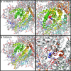
Lipid head groups may form part of the Cl conductance pathway in TMEM16A. a Molecular dynamics simulation of nhTMEM16 in a PtdCho bilayer, viewed from the extracellular space looking down the hydrophilic furrow formed by transmembrane helices α3, α4, α5, and α6. PtdCho molecules are shown in ball-and-stick representation colored by element (C=grey, blue=N, P=orange, O=red). Lipid head groups are seen in the furrow. Helices are numbered and colored as in Fig. 3. b–d Fantasy models of how ions may permeate TMEM16A and TMEM16F. b An homology model of TMEM16A was placed in register with nhTMEM16 using MatchMaker in UCSF Chimera with the Needleman-Wunsch alignment algorithm. The lipids were kept in the same absolute position as in A. The vestibule residues are red [129] and orange [86]. The Cl ion (green) was added to scale to show that it can fit between the lipid head groups and the protein. c Tricyanomethanide (C(CN3)) was positioned manually in the TMEM16A homology model. Functional residues are colored as in Fig. 4: the SCRD-homology domain [130] formed by α4 and α5 is red and the vestibule residues are orange. Although largely obscured by other regions of the protein, the selectivity filter is blue, the EEEEAVK sequence is magenta, and the Ca2+ binding residues are green. d NMDG+ was placed manually in the pore of the TMEM16F homology model
Fig. 7
Fig. 7
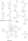
TMEM16A blockers are hydrophobic molecules. The structures include the classical TMEM16A blockers niflumic acid (NFA), anthacene-9-carboxylic acid (A9C), 4,4′-diisothiocyanostilbene-2,2′-disulfonic acid (DIDS), and 5-nitro-2-(3-phenylpropylamino)benzoic acid (NPPB) [38], and more recently identified inhibitors 1PBC [86], T16Ainh-A01 [79], benzbromarone [49], CaCCinh-A01 [26], MONNA [83], and tannic acid [80]. Other blockers not shown are also hydrophobic structures with aromatic rings

Similar articles

Cited by

References

    1. Adomaviciene A, Smith KJ, Garnett H, Tammaro P. Putative pore-loops of TMEM16/anoctamin channels affect channel density in cell membranes. J Physiol. 2013;591:3487–3505. doi: 10.1113/jphysiol.2013.251660. - DOI - PMC - PubMed
    1. Almaca J, Tian Y, Aldehni F, Ousingsawat J, Kongsuphol P, Rock JR, Harfe BD, Schreiber R, Kunzelmann K. TMEM16 proteins produce volume-regulated chloride currents that are reduced in mice lacking TMEM16A. J Biol Chem. 2009;284:28571–28578. doi: 10.1074/jbc.M109.010074. - DOI - PMC - PubMed
    1. Anderluh G, Dalla Serra M, Viero G, Guella G, Macek P, Menestrina G. Pore formation by equinatoxin II, a eukaryotic protein toxin, occurs by induction of nonlamellar lipid structures. J Biol Chem. 2003;278:45216–45223. doi: 10.1074/jbc.M305916200. - DOI - PubMed
    1. Andersen T, Haugen HK, Hotop H (1999) Binding energies in atomic negative ions: III. J Phys Chem Ref Data 28:1511–1533, doi:http://dx.doi.org/10.1063/1.556047 - DOI
    1. Anishkin A, Loukin SH, Teng J, Kung C. Feeling the hidden mechanical forces in lipid bilayer is an original sense. Proc Natl Acad Sci U S A. 2014;111:7898–7905. doi: 10.1073/pnas.1313364111. - DOI - PMC - PubMed

Publication types