. 2016 Feb 15;27(4):640-53.
doi: 10.1091/mbc.E15-03-0137. Epub 2016 Jan 6.

Evidence for new C-terminally truncated variants of α- and β-tubulins

Affiliations

Evidence for new C-terminally truncated variants of α- and β-tubulins

Chrystelle Aillaud et al. Mol Biol Cell. .

Abstract

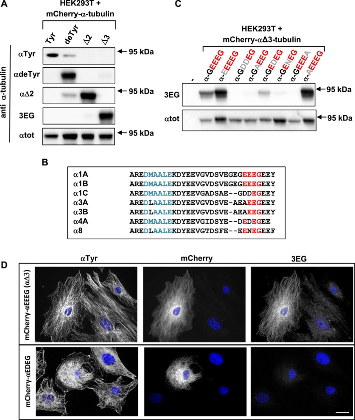
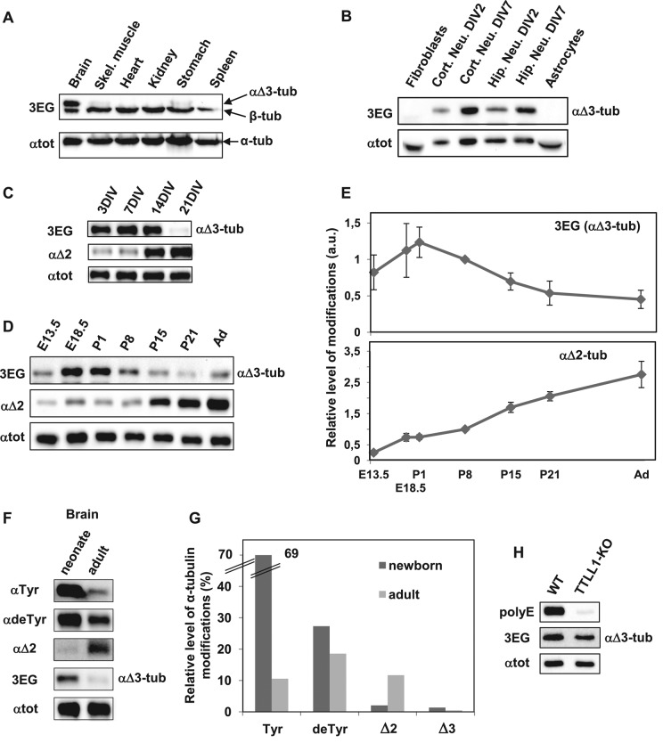
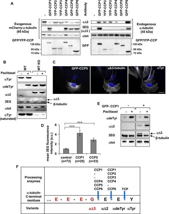
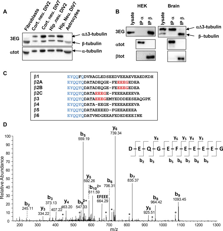
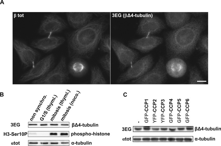
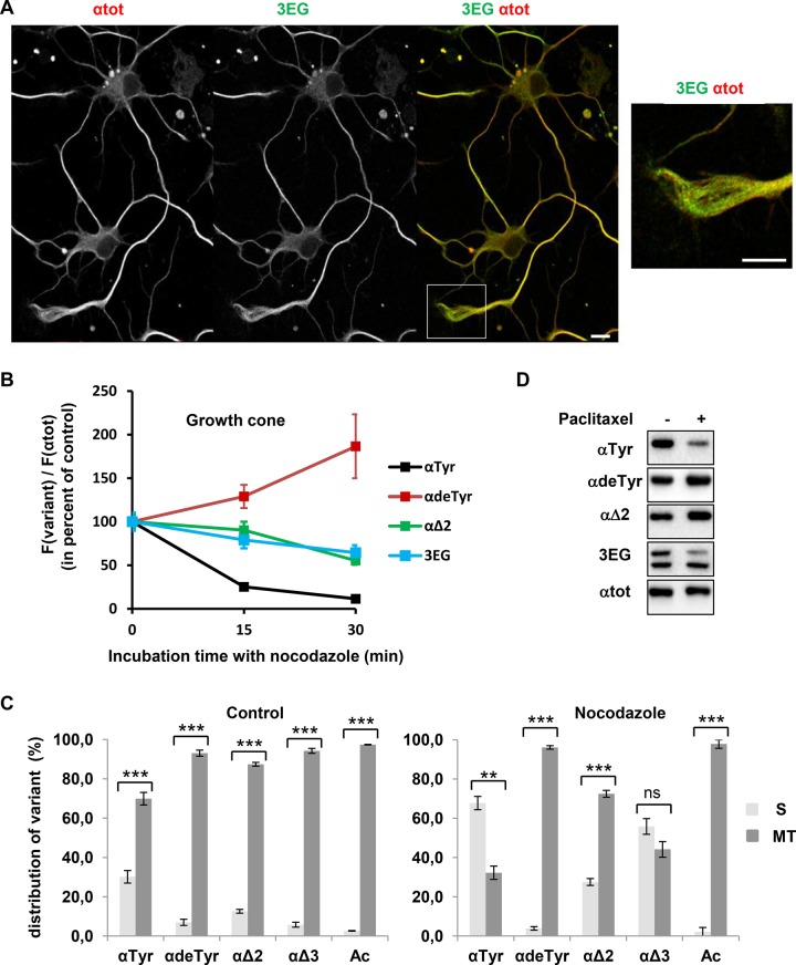
Cellular α-tubulin can bear various carboxy-terminal sequences: full-length tubulin arising from gene neosynthesis is tyrosinated, and two truncated variants, corresponding to detyrosinated and Δ2 α‑tubulin, result from the sequential cleavage of one or two C-terminal residues, respectively. Here, by using a novel antibody named 3EG that is highly specific to the -EEEG C-terminal sequence, we demonstrate the occurrence in neuronal tissues of a new αΔ3‑tubulin variant corresponding to α1A/B‑tubulin deleted of its last three residues (EEY). αΔ3‑tubulin has a specific distribution pattern: its quantity in the brain is similar to that of αΔ2-tubulin around birth but is much lower in adult tissue. This truncated α1A/B-tubulin variant can be generated from αΔ2-tubulin by the deglutamylases CCP1, CCP4, CCP5, and CCP6 but not by CCP2 and CCP3. Moreover, using 3EG antibody, we identify a C‑terminally truncated β-tubulin form with the same -EEEG C-terminal sequence. Using mass spectrometry, we demonstrate that β2A/B-tubulin is modified by truncation of the four C-terminal residues (EDEA). We show that this newly identified βΔ4-tubulin is ubiquitously present in cells and tissues and that its level is constant throughout the cell cycle. These new C-terminally truncated α- and β-tubulin variants, both ending with -EEEG sequence, are expected to regulate microtubule physiology. Of interest, the αΔ3-tubulin seems to be related to dynamic microtubules, resembling tyrosinated-tubulin rather than the other truncated variants, and may have critical function(s) in neuronal development.


Figures

FIGURE 1:
FIGURE 1:
A new antibody specific to the –EEEG protein C-terminus, named 3EG. (A, C) Immunoblot of protein extracts from HEK293T cells expressing different forms of 95-kDa α‑tubulin fused to mCherry at their N-terminus. Expression levels were controlled with αtot antibody. Antibodies recognizing either the unmodified C-terminal α-tubulin tail (tyrosinated tubulin) or its processed versions (detyrosinated or ∆2- or Δ3-tubulin) were used in A, and 3EG antibody was used in C to assay mutation (gray) of the Δ3-tubulin epitope (red). Quantification of the data in A is presented in Supplemental Table S1. (B) Alignment of mouse α-tubulin C-termini. Epitope of αtot antibody in blue; conserved amino acids from the –EEEG C-terminal sequence in red. (D) Immunocytochemistry of MEFs transfected with mCherry–αΔ3-tubulin and a mutated form of this protein ending with EDEG instead of EEEG. Scale bar: 20 µm.
FIGURE 2:
FIGURE 2:
αΔ3-Tubulin, a neuronal variant enriched around mouse birth. Equal quantities of proteins extracted from various tissues and cell types were subjected to immunoblot analysis. Protein levels were controlled using αtot antibody. (A) Immunoblot of crude protein extracts from the indicated neonate mouse tissues. (B) Immunoblot of protein extracts from the indicated cell types, including cortical and hippocampal neurons cultured 2 or 7 DIV. (C) Immunoblot of hippocampal neurons at different stages of culture. (D) Immunoblot of crude protein extracts from mouse brains at different stages of development, including E13.5 and E18.5, postnatal days 1, 8, 15 and 21 (P1–21), and adult (Ad). Mixes of four or five half-brains were used at each developmental stage. (E) Quantitative analysis of three immunoblots for αΔ3 and of five immunoblots for αΔ2 realized as in D with brain protein extracts. In each immunoblot, values obtained were normalized to the value obtained at P8. Error bars indicate SEM; a.u., arbitrary units. (F) Immunoblot of protein extracts from neonate (P1) and adult mouse brains (same extracts as in D). These samples were coanalyzed with extracts from HEK293T cells transfected with the various mCherry–α-tubulin variants (Supplemental Figure S2). (G) Quantitative analysis of immunoblots such as those presented in Supplemental Figure S2. Mixtures of five neonate brains and five adult half-brains were analyzed in two series of Western blots. The plotted values represent the percentages of the different forms of α-tubulin in brains estimated after normalization to total α-tubulin levels (with αtot antibody) and to antibody sensitivity (using modified mCherry–α-tubulins) as explained in Materials and Methods. (H) Immunoblot of crude protein extracts from wild-type (WT) and TTLL1-knockout mouse brains.
FIGURE 3:
FIGURE 3:
Specificity of CCP enzymes in producing αΔ2 and αΔ3. (A) Immunoblot of protein extracts from HEK293T cells coexpressing each GFP- or yellow fluorescent protein (YFP)–tagged CCP and mCherry–αΔ2-tubulin. Analysis of mCherry–α-tubulin (left) and endogenous α‑tubulin (right). (B) Immunoblot of protein extracted from WT and TTL-knockout (TTL KO) fibroblasts after incubation with dimethyl sulfoxide (DMSO: control) or paclitaxel (15 μM) for 2 h. TTL KO cells contain high levels of detyrosinated and αΔ2-tubulin, but no αΔ3-tubulin is detected. The reactive 3EG band corresponds to β-tubulin (Figure 4). (C) Immunocytochemistry of TTL KO fibroblasts transfected with GFP-CCP5 and immunostained with anti–αTyr-tubulin and 3EG antibody. Scale bar: 20 µm. CCP5 leads to the formation of αΔ3-tubulin. (D) Quantitative analysis of immunocytochemistry experiments as in C using TTL KO fibroblasts transfected with either GFP-CCP1 or GFP-CCP5. Fluorescence was measured as explained in Materials and Methods. (E) Immunoblot of protein extracted from HEK293T cells expressing GFP-CCP1 or not after incubation with DMSO (control) or paclitaxel (50 nM) for 24 h. (F) Schematic representation of the C-terminal amino acids of α1A/B-tubulin, 3EG epitope (red), and processing enzymes associated with the generation of the variants.
FIGURE 4:
FIGURE 4:
Evidence for a ubiquitous truncated form of β-tubulin. (A) Immunoblot of crude extracts from the indicated mouse tissues (same experiment as Figure 2B, but with the lower protein bands included). Protein levels were controlled using αtot antibody. (B) Immunoprecipitation of endogenous α- and β-tubulins from HEK293T cells and neonate mouse brain using αtot (IP α) and βtot (IP β) antibodies and analysis with 3EG antibody. The quality of immunoprecipitations was controlled using αtot and βtot antibodies together with mouse TrueBlot secondary antibodies. (C) Alignment of C-termini from the eight mouse β-tubulin isotypes. The βtot antibody epitope is in blue, and the 3EG epitope is in red. (D) Identification of the C-terminal peptide of the truncated β2A/B-tubulin (ΔEDEA) from neonatal mouse brain. The MS/MS spectrum of the peptide ion with m/z = 649.24, corresponding to an experimental monoprotonated mass MH+ of 1296.4632 Da, is annotated. The b- and y-type fragments identified are indicated on the sequence of the C-terminal peptide DEQGEFEEEEG, with a theoretical mass MH+ of 1296.4630 Da. Loss of H2O and NH3 is labeled with asterisks and open dots, respectively.
FIGURE 5:
FIGURE 5:
The novel β2A/B-tubulin variant βΔ4 evidenced in HeLa cells by 3EG antibody. (A) Immunocytochemistry of HeLa cells using 3EG and anti–β-tubulin antibody (βtot). Scale bar: 10 µm. (B) Immunoblot of protein extracts from nonsynchronized HeLa cells and from HeLa cells synchronized in G1/S or mitosis (see Materials and Methods). Cell synchronization was verified using a phosphohistone antibody (H3-Ser10P). Loaded proteins were standardized using αtot antibody, and the truncated β-tubulin form was analyzed with 3EG antibody. (C) Analysis of the β-tubulin band in protein extracts from HEK293T cells expressing GFP- or YFP-tagged CCPs (complementary analysis of the experiment in Figure 3A, right, including lower band).
FIGURE 6:
FIGURE 6:
Properties of neuronal 3EG-positive microtubules and tubulins. (A) Immunofluorescence study of the distribution of microtubules bearing αΔ3- and βΔ4-tubulin (3EG-positive microtubules) in hippocampal neurons cultured 2 DIV. Scale bar, 10 μm. Inset, magnification of the growth cone. Distribution of microtubules bearing the other α-tubulin variants is shown in Supplemental Figure S6. (B) Time course of nocodazole (20 μM) resistance of microtubules from 2 DIV hippocampal neurons analyzed in the growth cone (mean ± SEM). Microtubule fluorescence signals were measured for a minimum of 31 neurons at each time of drug treatment. The fluorescence signal of each tubulin variant, F(variant), was normalized to the total α-tubulin fluorescence signal, F(αtot), which is an index of the remaining microtubules, and then was plotted as percentage of the value obtained in the absence of nocodazole (time 0). (C) Immunoblot analysis of the distribution of α-tubulin species between soluble (S) and microtubular (MT) fractions in 7 DIV hippocampal neurons in the absence (left) or presence of 20 μM nocodazole for 30 min (right). The S and MT protein extracts from neuronal cultures (n = 3) were obtained as in Audebert et al. (1993). Results are shown as mean values ± SEM. ***p < 0.001 and **p < 0.01, t test. (D) Immunoblot of protein extracted from 7 DIV hippocampal neurons after incubation with DMSO (control) or paclitaxel (15 μM) for 2 h.

References

    1. Alexander JE, Hunt DF, Lee MK, Shabanowitz J, Michel H, Berlin SC, MacDonald TL, Sundberg RJ, Rebhun LI, Frankfurter A. Characterization of posttranslational modifications in neuron-specific class III beta-tubulin by mass spectrometry. Proc Natl Acad Sci USA. 1991;88:4685–4689. - PMC - PubMed
    1. Audebert S, Desbruyeres E, Gruszczynski C, Koulakoff A, Gros F, Denoulet P, Edde B. Reversible polyglutamylation of alpha- and beta-tubulin and microtubule dynamics in mouse brain neurons. Mol Biol Cell. 1993;4:615–626. - PMC - PubMed
    1. Baas PW, Ahmad FJ, Pienkowski TP, Brown A, Black MM. Sites of microtubule stabilization for the axon. J Neurosci. 1993;13:2177–2185. - PMC - PubMed
    1. Badin-Larcon AC, Boscheron C, Soleilhac JM, Piel M, Mann C, Denarier E, Fourest-Lieuvin A, Lafanechere L, Bornens M, Job D. Suppression of nuclear oscillations in Saccharomyces cerevisiae expressing Glu tubulin. Proc Natl Acad Sci USA. 2004;101:5577–5582. - PMC - PubMed
    1. Berezniuk I, Lyons PJ, Sironi JJ, Xiao H, Setou M, Angeletti RH, Ikegami K, Fricker LD. Cytosolic carboxypeptidase 5 removes alpha- and gamma-linked glutamates from tubulin. J Biol Chem. 2013;288:30445–30453. - PMC - PubMed

Publication types

MeSH terms