

Arsenic-induced Histological Alterations in Various Organs of Mice
- PMID: 26740907
- PMCID: PMC4698904
- DOI: 10.4172/2157-7099.1000323
Arsenic-induced Histological Alterations in Various Organs of Mice
Abstract
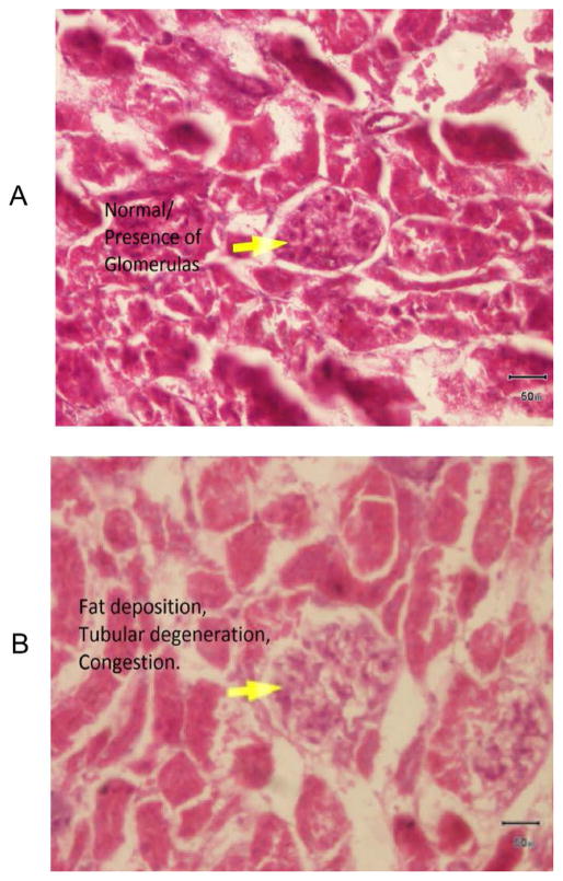
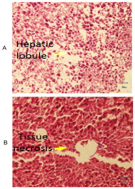
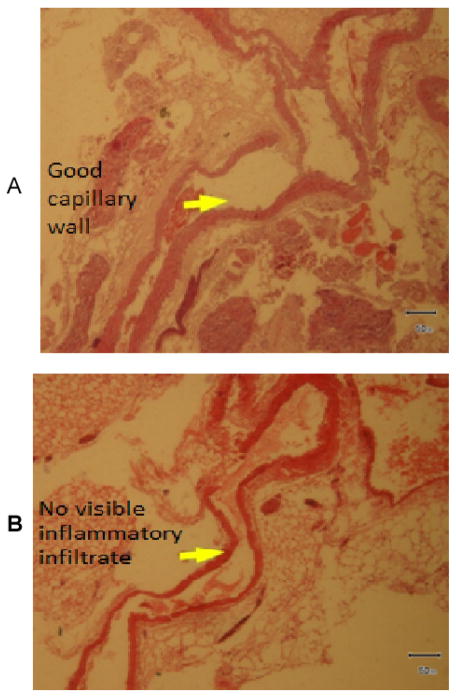
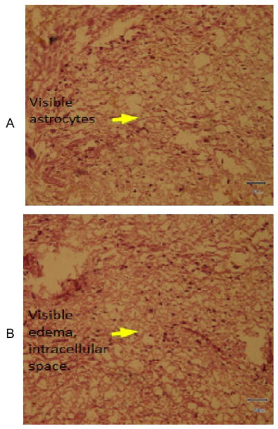
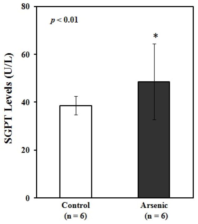
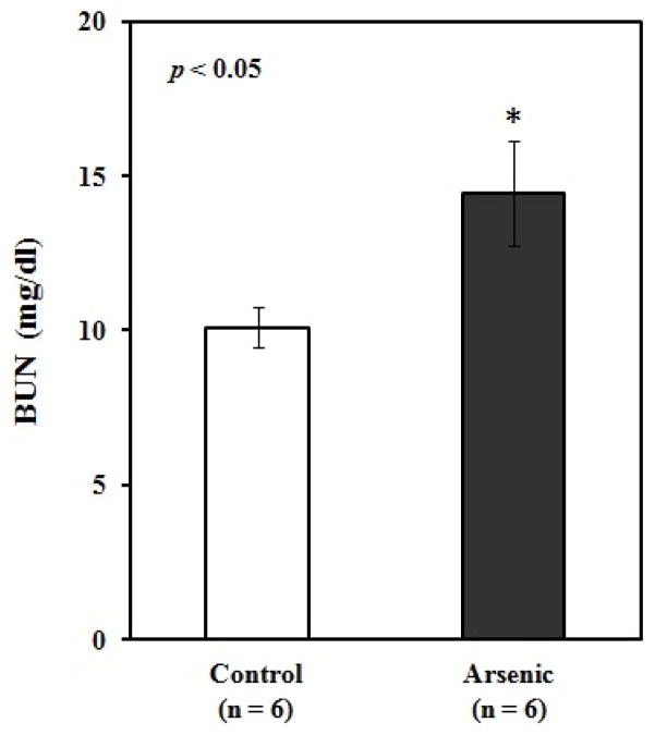
Deposition of arsenic in mice through groundwater is well documented but little is known about the histological changes of organs by the metalloid. Present study was designed to evaluate arsenic-induced histological alterations in kidney, liver, thoracic artery and brain of mice which are not well documented yet. Swiss albino male mice were divided into 2 groups and treated as follows: Group 1: control, 2: arsenic (sodium arsenite at 10 mg/kg b.w. orally for 8 wks). Group 2 showed marked degenerative changes in kidney, liver, thoracic artery, and brain whereas Group 1 did not reveal any abnormalities on histopathology. We therefore concluded that arsenic induces histological alterations in the tested organs.
Keywords: Arsenic; Histopathology; Mice organs.
Figures
References
-
- Ravenscroft P, Brammer H, Richards KS. Arsenic Pollution: A Global Synthesis. 1. Wiley-Blackwell; UK: 2009.
-
- Tseng WP, Chu HM, How SW, Fong JM, Lin CS, et al. Prevalence of skin cancer in an endemic area of chronic arsenicism in Taiwan. J Natl Cancer Inst. 1968;40:453–463. - PubMed
-
- Cebrian ME, Albores A, Aguilar M, Blakely E. Chronic arsenic poisoning in the north of Mexico. Hum Toxicol. 1983;2:121–133. - PubMed
-
- Vahidnia A, van der Straaten RJ, Romijn F, van Pelt J, van der Voet GB, et al. Arsenic metabolites affect expression of the neurofilament and tau genes: an in-vitro study into the mechanism of arsenic neurotoxicity. Toxicol In Vitro. 2007;2:1104–1112. - PubMed
-
- Jolliffe DM, Budd AJ, Gwilt DJ. Massive acute arsenic poisoning. Anesthesia. 1991;46:288–290. - PubMed
Grants and funding
LinkOut - more resources
Full Text Sources
Other Literature Sources