. 2015 Dec 31;4(6):8.
doi: 10.1167/tvst.4.6.8. eCollection 2015 Dec.

Tunnel Vision Prismatic Field Expansion: Challenges and Requirements

Affiliations

Tunnel Vision Prismatic Field Expansion: Challenges and Requirements

Henry Apfelbaum et al. Transl Vis Sci Technol. .

Abstract

Purpose: No prismatic solution for peripheral field loss (PFL) has gained widespread acceptance. Field extended by prisms has a corresponding optical scotoma at the prism apices. True expansion can be achieved when each eye is given a different view (through visual confusion). We analyze the effects of apical scotomas and binocular visual confusion in different designs to identify constraints on any solution that is likely to meet acceptance.

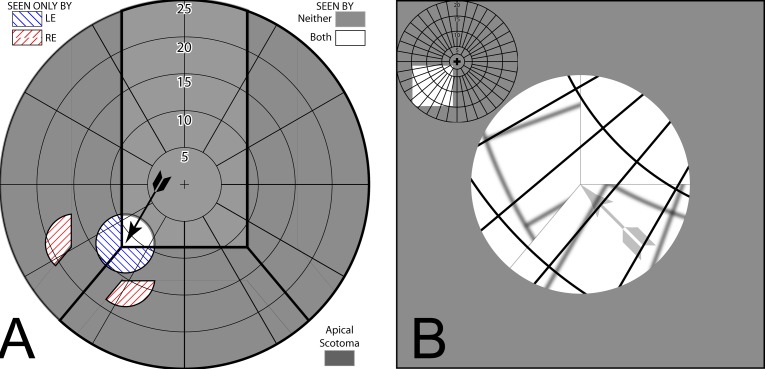
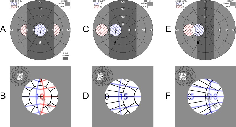
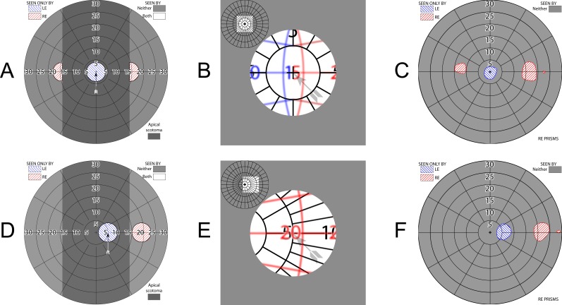
Methods: Calculated perimetry diagrams were compared to perimetry with PFL patients wearing InWave channel prisms and Trifield spectacles. Percept diagrams illustrate the binocular visual confusion.

Results: Channel prisms provide no benefit at primary gaze. Inconsequential extension was provided by InWave prisms, although accessible with moderate gaze shifts. Higher-power prisms provide greater extension, with greater paracentral scotoma loss, but require uncomfortable gaze shifts. Head turns, not eye scans, are needed to see regions lost to the apical scotomas. Trifield prisms provide field expansion at all gaze positions, but acceptance was limited by disturbing effects of central binocular visual confusion.

Conclusions: Field expansion when at primary gaze (where most time is spent) is needed while still providing unobstructed central vision. Paracentral multiplexing prisms we are developing that superimpose shifted and see-through views may accomplish that.

Translational relevance: Use of the analyses and diagramming techniques presented here will be of value when considering prismatic aids for PFL, and could have prevented many unsuccessful designs and the improbable reports we cited from the literature. New designs must likely address the challenges identified here.

Keywords: low vision rehabilitation; perimetry; prism treatment; retinitis pigmentosa; tunnel vision; visual field loss.


Figures

Figure 1
Figure 1
Visual confusion and diplopia. (A) A savannah cartoon. The blue and red dashed outlines represent the field of view of the left eye (LE) and right eye (RE), respectively, when a 20° shift is provided by a full-lens base-out prism (∼36Δ) in front of the LE. The red cross (not part of the scene) marks the fixation location of the nonprism RE. (B) The way this scene appears in the binocular visual field of an observer with no visual field loss. Everything within the intersection of the blue and red outlines in (A) is seen diplopically (i.e., at two apparent locations), while everything within the intersection of the red and blue outlines in (b) is seen with visual confusion (superposition of different images at the same apparent direction). Thus the LE view of the lion overlaps the RE view of the cage, the LE view of the cage has captured the RE view of the cub, and the giraffe is now prey for the lion and tiger. The LE is bathing the cub in the RE's pond, but the hippo is no closer to bathing in the pond since both pond and hippo are only seen by the RE. Thus the pond is seen in confusion but not diplopia, while the hippo and rhino are seen without confusion or diplopia. Although the tiger is seen in a region with visual confusion, there happens to be no conflicting RE salient image at that location. Visual confusion is primarily a problem only when salient images appear in both eyes at corresponding retinal locations (directions), and is more disturbing centrally than in the periphery. Diplopia is always noticeable, but more disturbing centrally than in the periphery. (C) Calculated dichoptic perimetry for this configuration. By convention, the diagram is limited to a radius of 90°, as is a Goldmann perimeter, although the field of view in this case actually extends left to 110°. Note the direct relationship between (A) and (C), with everything in the central white area of (C) seen diplopically. (D) The corresponding percept diagram identifies visual confusion, as shown by the superimposed images representing the view of a perimetry grid seen by each eye. LE views are blurred slightly to distinguish them and represent the loss of optical quality through the prism, and the light gray arrow (not part of the patient's percept) points to fixation in these percept diagrams. Note the direct relationship between (B) and (D). (E) Calculated perimetry for a patient with 20° residual visual fields and the same prism configuration. Since entirely different portions of the scene are viewed by each eye, there is no diplopia. (F) The corresponding percept diagram (with just the central portion of interest shown). There is visual confusion everywhere. Thus with PFL, confusion without diplopia is possible, allowing for field of view expansion without diplopia. (Diplopia without visual confusion is a possibility in cases of bitemporal or binasal hemianopia with phorias.)
Figure 2
Figure 2
Channel prisms. (A) Channel prism lens available from Chadwick Optical. There are three sector prisms per carrier lens, with the apices outside the borders of the patient's functional field when at primary gaze. In this case, the lateral prisms are 12Δ, base-in and base-out, on the nasal and temporal side, respectively, and the lower prisms are 8Δ, base-down, matching the configuration and prism powers that were used by InWave. Note the break in the visibility of the spectacles' temples due to the apical scotomas. (B) Photo through a molded InWave channel prism trial lens. A polar grid with 5° radius increments is seen through the lens. The channel width is 6 mm (17°), the smallest offered by InWave. Note that the central circle in the grid is 10° in diameter. Approximately 6° laterally are lost to each of the left and right prisms' apical scotomas to achieve a corresponding lateral field substitution (extension, not expansion), and 4° lost to the lower scotoma, hiding sections of the original grid from view. (C) 6 mm channel lens fabricated using 40Δ 3M Press-On™ Fresnel prisms. High-power prisms have correspondingly large apical scotomas. (The spectacles are just a few millimeters from the paper, yet the apical scotoma changes each fox into an ox.)
Figure 3
Figure 3
Trifield prism spectacles. (A) Front view. (B) View from above. Tinted prisms aid in determining veridical direction of objects seen through them.
Figure 4
Figure 4
InWave channel prism fields. (A) Calculated perimetry with 8° residual visual field at primary gaze in a 6-mm channel. The prisms have no effect. Field of view shaded darker is lost to the apical scotomas. (B) With gaze shifted 8.5° (of visual field) left and down to the channel corner (9° of diagonal ocular rotation), the field is split into three parts, giving a 6° extension left and 4° down, with corresponding gaps in between at the apical scotomas. (C) The percept diagram corresponding to (B) for this bilateral fitting shows the larger grid spacing of the shifted fields and perceived discontinuity (jump) in the field of view caused by the scotomas. Thin gray lines, not part of the patient's percept, are shown to identify the prism apices. There is no visual confusion or diplopia, nor field expansion. (D) Measured LE field of patient 1 at this gaze position (RE patched). The total area seen is slightly larger than the 8° residual field due to vignetting at the prism edges. Dashed lines indicate the apparent location of the prism apices, a bit to the right of the intended location. (E) The asymmetric monocular field of patient 2 required shifting the 12 mm (33°) channel to the right, so that the residual field at primary gaze lies entirely within the channel. (F) With fixation shifted 7° left and 9° degrees down, the channel corner splits the field. Even though the channel and residual field sizes differ greatly for these two patients, the extension provided is the same. The apparent difference in scotoma sizes is an artifact of the imprecision of these difficult measurements, also seen as the differences in field size and shape between (E) and (F) for the 20/500 acuity of this subject.
Figure 5
Figure 5
Unilateral fitting. (A) With a channel prism lens fit only on the right eye (RE), the RE prism views provide true field expansion, as the LE sees the regions lost to the RE apical scotomas. (B) The percept diagram shows that the expansion comes at the expense of visual confusion.
Figure 6
Figure 6
Trifield prisms. (A) Simulated perimetry at primary gaze. The two 16Δ prisms add two peripheral islands of visibility. Two adjoining (but only monocular) apical scotomas, each larger than the patient's 8° field width, are evident (darker gray shading). (B) Corresponding percept diagram. Blue and red tints in the percept diagrams identify field viewed through the tinted right eye (RE) left (green) and right (red) prisms, respectively. (Although the prisms are tinted red and green, we diagram with red and blue in deference to dichromats.) Left eye (LE) maintains a direct, nonprismatic view at all gaze positions. True field expansion comes at the expense of central visual confusion. (C) With a left gaze shift of about half the residual field width, the RE view is entirely within the left green prism and a relatively continuous expanded view is afforded, without diplopia. (D) The corresponding percept diagram shows that there is still central confusion, but only from two views, not three. The digit 5 is coincidentally overlaid. (E) With 10Δ prisms, the power is not sufficient to avoid undesirable diplopia. (F) A region including the zero of the 10° eccentricity marking is thus perceived diplopically.
Figure 7
Figure 7
Patient 1 Trifield results. The patient's fields have shrunk in the decade since the 25Δ prisms were fitted, resulting in larger field of view gaps than illustrated in Figure 6A (and the diagram scale has changed to accommodate the higher-power prisms). Unlike channel prisms, which provide no access to the scotoma areas even with gaze shifts, the fellow nonprism eye can see into the apical scotoma regions, so the loss (without head turning) is not as problematic. Having access at primary gaze to the region at eccentricities larger than the scotomas may be desirable, although that has not been studied. (A) Simulated perimetry at primary gaze. (B) Corresponding percept diagram shows the “Trifield” binocular central visual confusion. (C) Corresponding perimetry. The asymmetry is indicative of the difficulty of positioning the prism apices exactly at primary gaze, as the perimeter's fixation target is invisible at that location in the prisms, and the eyes readily dissociate into the phoria posture, without any clue for the directional shift needed to align the eyes to assess veridical direction. (However, since the prisms are intended to provide hazard detection followed by a gaze turn to foveate the hazard with the nonprism eye, precise angular perception may not be needed.) (D) With a 5° gaze shift toward the right, the right eye (RE) view is entirely through the right prism. (E) The corresponding percept diagram again illustrates that there is still central confusion showing, but only from two views, not three. (F) Corresponding perimetry. We use the symbology of our dichoptic perimeter, but dichoptic shutter goggles were not needed to identify each eye's obvious and separate contribution.

Similar articles

Cited by

References

    1. Geruschat DR,, Turano KA. Connecting research on retinitis pigmentosa to the practice of orientation and mobility. J Vis Impair Blind. 2002; 96; 69–85.
    1. Drasdo N. Techniques instruments, cases; visual field expanders. Am J Optom Physiol Opt. 1976; 53 (9 pt 1): 464–467. - PubMed
    1. Krefman RA. Reversed telescopes on visual efficiency scores in field-restricted patients. Am J Optom Physiol Opt. 1981; 58; 159–162. - PubMed
    1. Hoeft WW,, Feinbloom W,, Brilliant R,, et al. Amorphic lenses: a mobility aid for patients with retinitis pigmentosa. Am J Optom Physiol Opt. 1985; 62: 142–148. - PubMed
    1. Szlyk JP,, Seiple W,, Laderman DJ,, Kelsch R,, Ho K,, McMahon T. Use of bioptic amorphic lenses to expand the visual field in patients with peripheral loss. Optom Vis Sci. 1998; 75; 518–524. - PubMed

LinkOut - more resources