

Automatic segmentation of the hippocampus for preterm neonates from early-in-life to term-equivalent age
- PMID: 26740912
- PMCID: PMC4561668
- DOI: 10.1016/j.nicl.2015.07.019
Automatic segmentation of the hippocampus for preterm neonates from early-in-life to term-equivalent age
Abstract
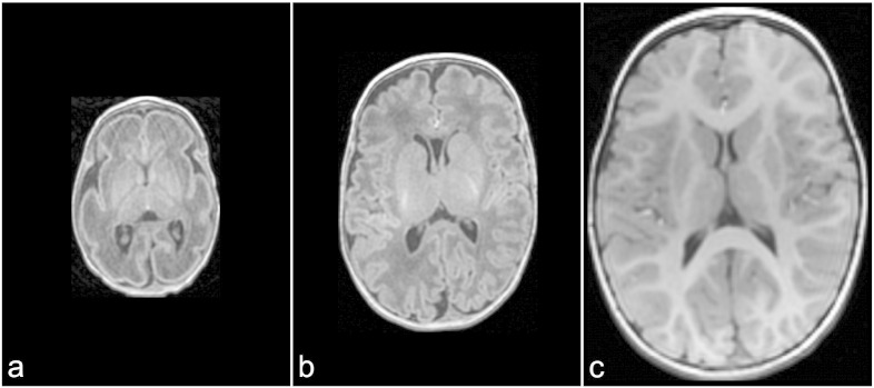
Introduction: The hippocampus, a medial temporal lobe structure central to learning and memory, is particularly vulnerable in preterm-born neonates. To date, segmentation of the hippocampus for preterm-born neonates has not yet been performed early-in-life (shortly after birth when clinically stable). The present study focuses on the development and validation of an automatic segmentation protocol that is based on the MAGeT-Brain (Multiple Automatically Generated Templates) algorithm to delineate the hippocampi of preterm neonates on their brain MRIs acquired at not only term-equivalent age but also early-in-life.
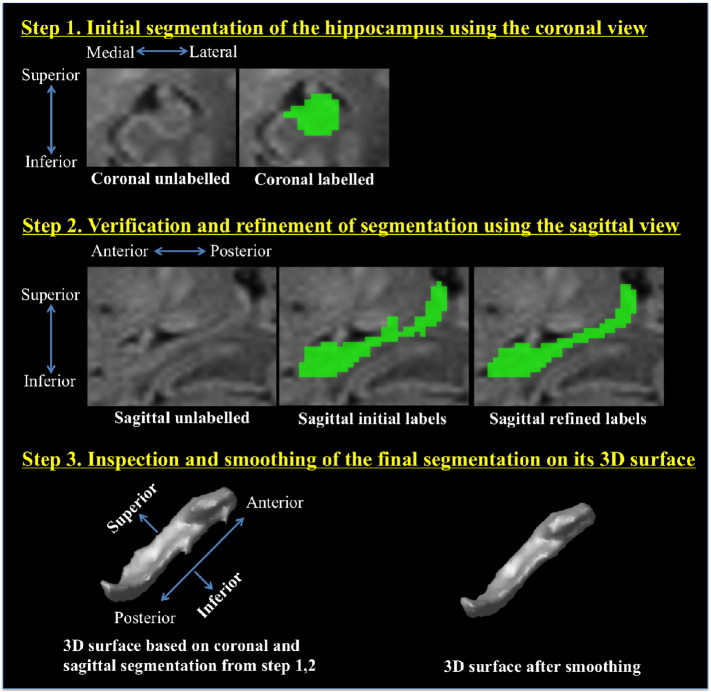
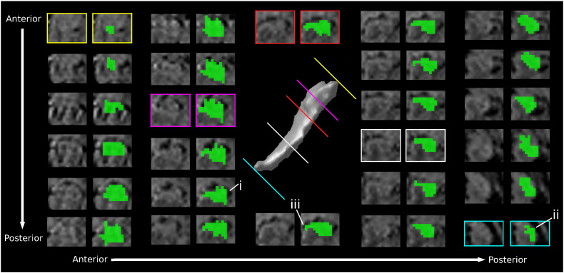
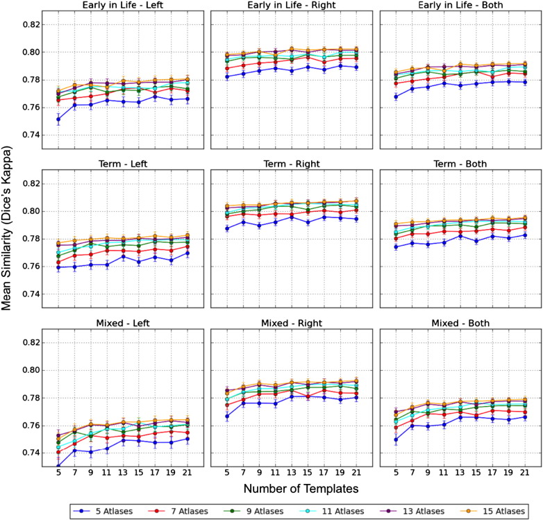
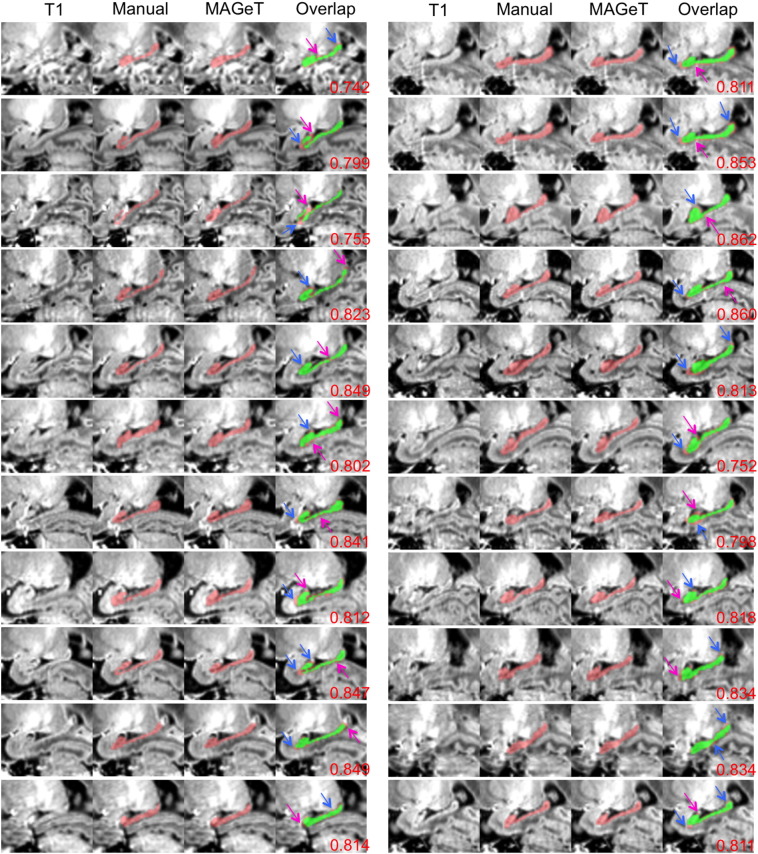
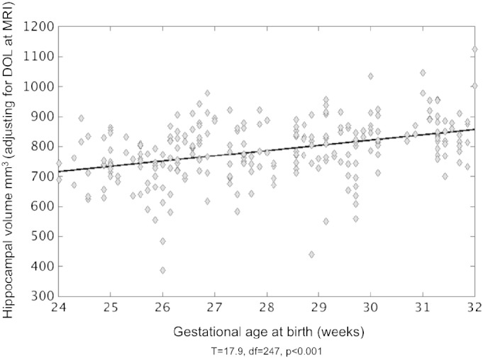
Methods: First, we present a three-step manual segmentation protocol to delineate the hippocampus for preterm neonates and apply this protocol on 22 early-in-life and 22 term images. These manual segmentations are considered the gold standard in assessing the automatic segmentations. MAGeT-Brain, automatic hippocampal segmentation pipeline, requires only a small number of input atlases and reduces the registration and resampling errors by employing an intermediate template library. We assess the segmentation accuracy of MAGeT-Brain in three validation studies, evaluate the hippocampal growth from early-in-life to term-equivalent age, and study the effect of preterm birth on the hippocampal volume. The first experiment thoroughly validates MAGeT-Brain segmentation in three sets of 10-fold Monte Carlo cross-validation (MCCV) analyses with 187 different groups of input atlases and templates. The second experiment segments the neonatal hippocampi on 168 early-in-life and 154 term images and evaluates the hippocampal growth rate of 125 infants from early-in-life to term-equivalent age. The third experiment analyzes the effect of gestational age (GA) at birth on the average hippocampal volume at early-in-life and term-equivalent age using linear regression.
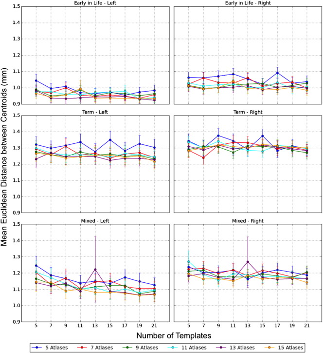
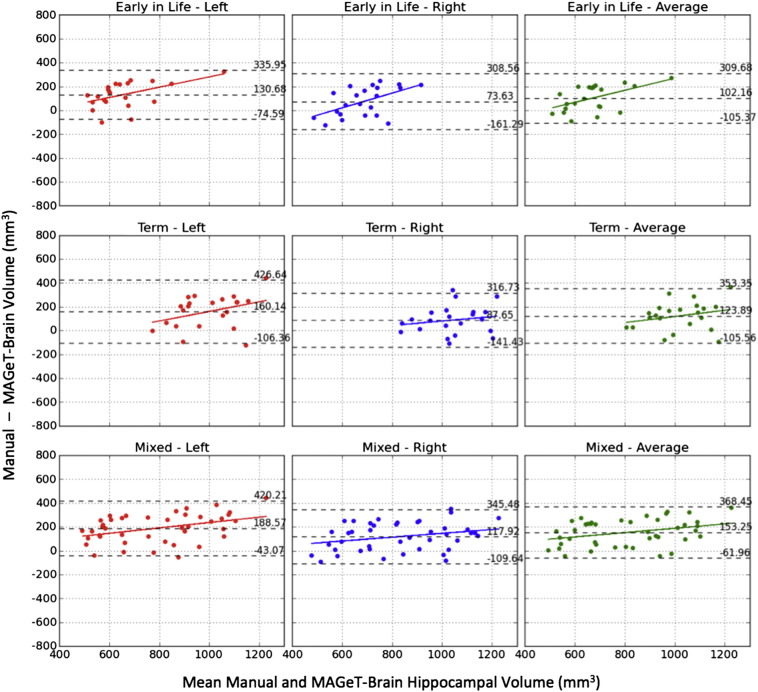
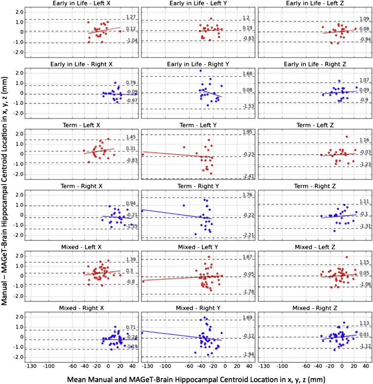
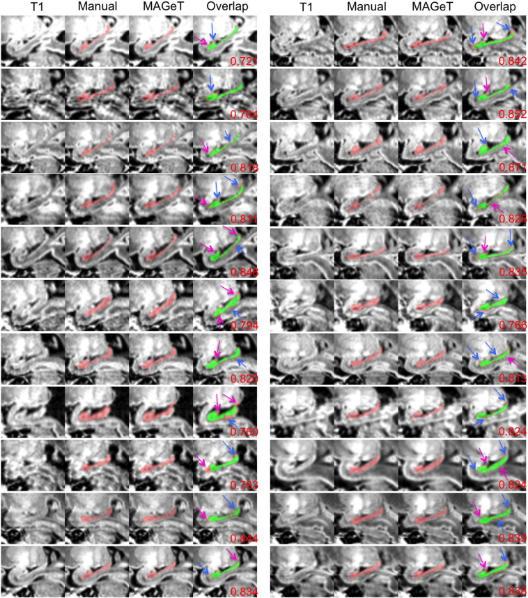
Results: The final segmentations demonstrate that MAGeT-Brain consistently provides accurate segmentations in comparison to manually derived gold standards (mean Dice's Kappa > 0.79 and Euclidean distance <1.3 mm between centroids). Using this method, we demonstrate that the average volume of the hippocampus is significantly different (p < 0.0001) in early-in-life (621.8 mm(3)) and term-equivalent age (958.8 mm(3)). Using these differences, we generalize the hippocampal growth rate to 38.3 ± 11.7 mm(3)/week and 40.5 ± 12.9 mm(3)/week for the left and right hippocampi respectively. Not surprisingly, younger gestational age at birth is associated with smaller volumes of the hippocampi (p = 0.001).
Conclusions: MAGeT-Brain is capable of segmenting hippocampi accurately in preterm neonates, even at early-in-life. Hippocampal asymmetry with a larger right side is demonstrated on early-in-life images, suggesting that this phenomenon has its onset in the 3rd trimester of gestation. Hippocampal volume assessed at the time of early-in-life and term-equivalent age is linearly associated with GA at birth, whereby smaller volumes are associated with earlier birth.
Keywords: Early-in-life; Hippocampus; MAGeT-Brain; MRI; Preterm neonates; Segmentation.
Figures
Similar articles
-
Multi-atlas segmentation of the whole hippocampus and subfields using multiple automatically generated templates.Neuroimage. 2014 Nov 1;101:494-512. doi: 10.1016/j.neuroimage.2014.04.054. Epub 2014 Apr 29. Neuroimage. 2014. PMID: 24784800
-
Accuracy and bias of automatic hippocampal segmentation in children and adolescents.Brain Struct Funct. 2019 Mar;224(2):795-810. doi: 10.1007/s00429-018-1802-2. Epub 2018 Dec 3. Brain Struct Funct. 2019. PMID: 30511334
-
Training labels for hippocampal segmentation based on the EADC-ADNI harmonized hippocampal protocol.Alzheimers Dement. 2015 Feb;11(2):175-83. doi: 10.1016/j.jalz.2014.12.002. Epub 2015 Jan 20. Alzheimers Dement. 2015. PMID: 25616957
-
Meta-Analysis of Hippocampal Volume and Episodic Memory in Preterm and Term Born Individuals.Neuropsychol Rev. 2024 Jun;34(2):478-495. doi: 10.1007/s11065-023-09583-6. Epub 2023 Apr 15. Neuropsychol Rev. 2024. PMID: 37060422 Review.
-
Hippocampal subfields at ultra high field MRI: An overview of segmentation and measurement methods.Hippocampus. 2017 May;27(5):481-494. doi: 10.1002/hipo.22717. Epub 2017 Feb 23. Hippocampus. 2017. PMID: 28188659 Free PMC article. Review.
Cited by
-
Early Procedural Pain Is Associated with Regionally-Specific Alterations in Thalamic Development in Preterm Neonates.J Neurosci. 2018 Jan 24;38(4):878-886. doi: 10.1523/JNEUROSCI.0867-17.2017. Epub 2017 Dec 18. J Neurosci. 2018. PMID: 29255007 Free PMC article.
-
MRI based radiomics enhances prediction of neurodevelopmental outcome in very preterm neonates.Sci Rep. 2022 Jul 13;12(1):11872. doi: 10.1038/s41598-022-16066-w. Sci Rep. 2022. PMID: 35831452 Free PMC article.
-
Hippocampus, Amygdala, and Thalamus Volumes in Very Preterm Children at 8 Years: Neonatal Pain and Genetic Variation.Front Behav Neurosci. 2019 Mar 19;13:51. doi: 10.3389/fnbeh.2019.00051. eCollection 2019. Front Behav Neurosci. 2019. PMID: 30941021 Free PMC article.
-
Polygenic risk for depression and anterior and posterior hippocampal volume in children and adolescents.J Affect Disord. 2024 Jan 1;344:619-627. doi: 10.1016/j.jad.2023.10.068. Epub 2023 Oct 18. J Affect Disord. 2024. PMID: 37858734 Free PMC article.
-
Children's family income is associated with cognitive function and volume of anterior not posterior hippocampus.Nat Commun. 2020 Aug 12;11(1):4040. doi: 10.1038/s41467-020-17854-6. Nat Commun. 2020. PMID: 32788583 Free PMC article.
References
Publication types
MeSH terms
Grants and funding
LinkOut - more resources
Full Text Sources
Other Literature Sources
Medical
