. 2016 Apr 15;11(4):992-1000.
doi: 10.1021/acschembio.5b00806. Epub 2016 Jan 15.

Stable Colloidal Drug Aggregates Catch and Release Active Enzymes

Affiliations

Stable Colloidal Drug Aggregates Catch and Release Active Enzymes

Christopher K McLaughlin et al. ACS Chem Biol. .

Abstract

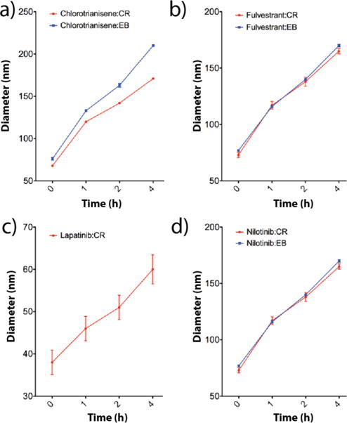
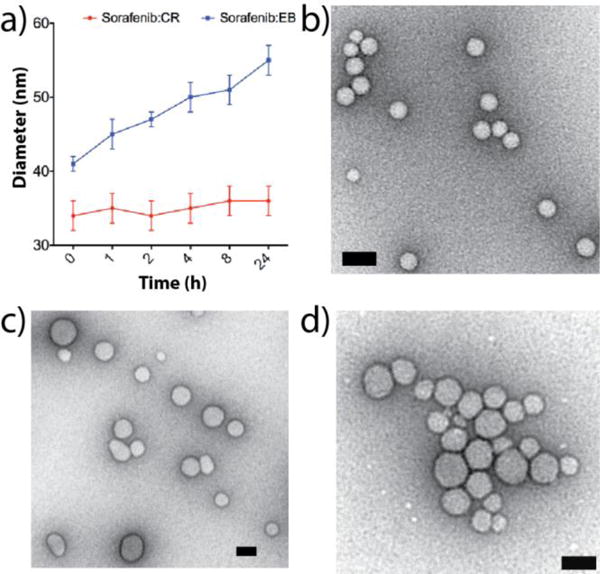
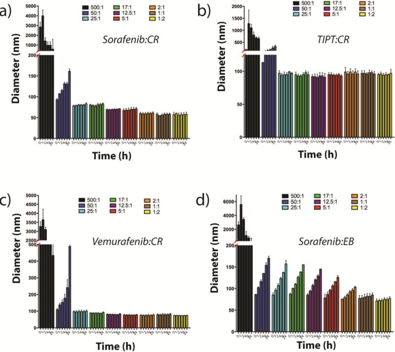
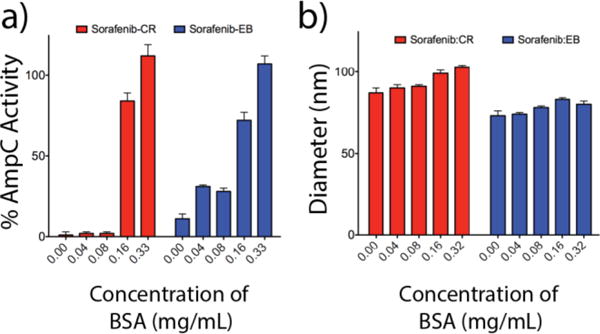
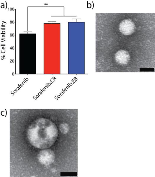
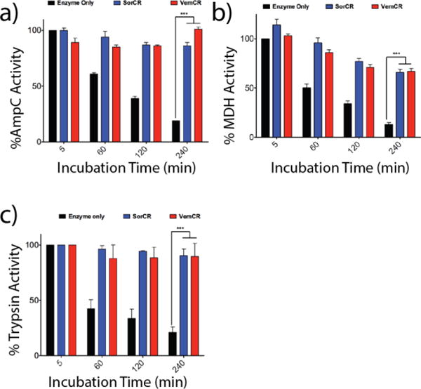
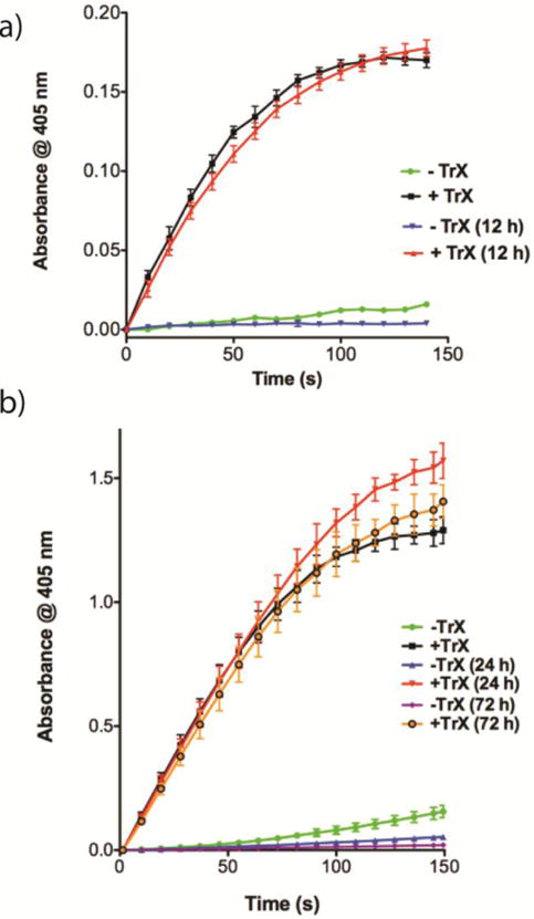
Small molecule aggregates are considered nuisance compounds in drug discovery, but their unusual properties as colloids could be exploited to form stable vehicles to preserve protein activity. We investigated the coaggregation of seven molecules chosen because they had been previously intensely studied as colloidal aggregators, coformulating them with bis-azo dyes. The coformulation reduced colloid sizes to <100 nm and improved uniformity of the particle size distribution. The new colloid formulations are more stable than previous aggregator particles. Specifically, coaggregation of Congo Red with sorafenib, tetraiodophenolphthalein (TIPT), or vemurafenib produced particles that are stable in solutions of high ionic strength and high protein concentrations. Like traditional, single compound colloidal aggregates, the stabilized colloids adsorbed and inhibited enzymes like β-lactamase, malate dehydrogenase, and trypsin. Unlike traditional aggregates, the coformulated colloid-protein particles could be centrifuged and resuspended multiple times, and from resuspended particles, active trypsin could be released up to 72 h after adsorption. Unexpectedly, the stable colloidal formulations can sequester, stabilize, and isolate enzymes by spin-down, resuspension, and release.


Conflict of interest statement

Notes The authors declare no competing financial interest.

Figures

Figure 1
Figure 1
Colloidal formulations containing 50 uM drug and 50 uM of either CR or EB all increase in diameter after 4 h, as measured by DLS: a) chlorotrianisene:CR and chlorotrianisene:EB, b) fulvestrant:CR and fulvestrant:EB, c) lapatinib:CR and d) nilotinib:CR and nilotinib:EB (n=3 independent colloid formulations, mean± SD). Lapatinib:EB is not included as this solution precipitated within 1 h.
Figure 2
Figure 2
a) Sorafenib:CR (red line) and sorafenib:EB (50 μM per compound, PBS) show little change in hydrodynamic diameters over a 24 h incubation period (n = 3 independent colloid formulations, mean ± SD). b–d) TEM images for b) sorafenib:CR, c) TIPT:CR, and d) vemurafenib:CR reveal homogeneous populations of particles (scale bars 50 nm).
Figure 3
Figure 3
DLS measurements taken over a 24 h incubation period for a) sorafenib:CR, b) TIPT:CR, c) sorafenib:CR, and d) sorafenib:EB reveal changes in particle diameters based on the concentration of dye used. For all CR formulated colloids (a–c), excellent sizing and stability are achieved starting at a drug:dye ratio of 25:1 (drug:dye = 500 μM:X, where X = 1, 10, 20, 30, 40, 100, 250, 500 and 1000 μM; n = 3 independent colloid formulations, mean ± SD).
Figure 4
Figure 4
Effect of BSA on enzyme activity. a) Inhibition data for sorafenib:CR and sorafenib:EB (1:1, 100 μM, KPi) with varying concentrations of BSA obtained using the enzyme AmpC β-lactamase (n=3, mean ± SD). Full enzyme activity is recovered upon dilution into 0.32 mg/mL BSA for both formulations. b) Corresponding hydrodynamic diameters for each of the enzyme inhibition data points reveal particle stability despite the adsorption of protein.
Figure 5
Figure 5
a) In vitro assessment of co-aggregated formulations sorafenib:CR (red bars) and sorafenib:EB (blue bars, 1:1, 50 μM each) with MDA-MB-231 cells reveals significantly less cytotoxicity compared to sorafenib colloids alone (black bars) (n = 3 independent measurements, mean ± SD, ** denotes p < 0.01). Representative TEM fields of view for b) sorafenib:CR and c) sorafenib:EB (1:1, 50 μM each, PBS) obtained after incubation in BSA (4 mg/mL, PBS, 4h, 37 °C). Scale bars are 50 nm.
Figure 6
Figure 6
Incubation of enzyme alone (black bars), sorafenib:CR (blue bars) and vemurafenib:CR (red bars) with enzymes (a) AmpC, (b) MDH and (c) trypsin reveal significant differences in activity over a 4 h incubation period (n = 3, mean ± SD ***p < 0.001). For all three enzymes, activity is drastically reduced for the enzyme only samples with time. Adsorption to either colloidal formulation followed by subsequent particle disruption using Triton X-100 (0.1%) restored enzyme activity.
Figure 7
Figure 7
a) Centrifugation after sorafenib:CR (25:1, 500 μM, KPi) incubation with AmpC resulted in a red pellet that could be easily re-suspended in KPi buffer (n = 3, mean ± SD). Addition of β-lactamase substrate CENTA™ substrate led to limited substrate cleavage (green line). Colloid disruption using 0.1% TrX restored activity and resulted in substrate cleavage as indicated by the increase in absorbance at 405 nm (black line). Colloid-enzyme complexes were left for 12 h, after which nearly identical activity was found for solutions without (blue line) and with (red line) TrX addition. b) Studies with trypsin revealed similar trends as outlined above (n = 3, mean ± SD). Activity here was monitored after 0, 24 and 72 h. For both a) and b), activity of the released enzymes between 0 and 12, 24 or 72 h is not statistically significant.

References

    1. Shoichet BK. Screening in a spirit haunted world. Drug discovery today. 2006;11:607–615. - PMC - PubMed
    1. Brick MC, Palmer HJ, Whitesides TH. Formation of colloidal dispersions of organic materials in aqueous media by solvent shifting. Langmuir. 2003;19:6367–6380.
    1. McGovern SL, Caselli E, Grigorieff N, Shoichet BK. A common mechanism underlying promiscuous inhibitors from virtual and high-throughput screening. J Med Chem. 2002;45:1712–1722. - PubMed
    1. McGovern SL, Helfand BT, Feng B, Shoichet BK. A specific mechanism of nonspecific inhibition. J Med Chem. 2003;46:4265–4272. - PubMed
    1. Thorne N, Auld DS, Inglese J. Apparent activity in high-throughput screening: origins of compound-dependent assay interference. Curr Opin Chem Biol. 2010;14:315–324. - PMC - PubMed

Publication types

MeSH terms